What to Expect with Pterygium Surgery

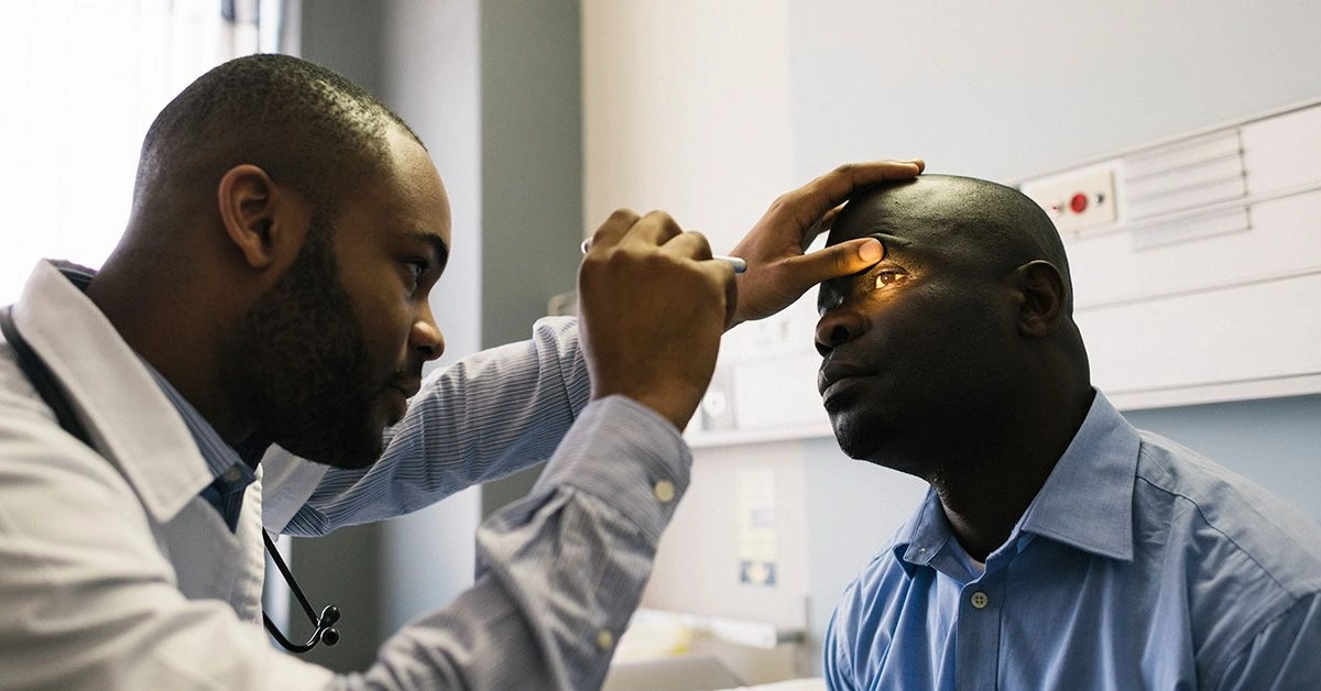
Overview
Pterygium surgery is performed to excise noncancerous growths of the conjunctiva (pterygia) from the eye.
The conjunctiva is the transparent tissue that covers the sclera (the white of the eye) and the inner surface of the eyelids. Some pterygia cause minimal or no symptoms. In more advanced cases, excessive conjunctival growth can extend onto the cornea and impair vision.
Presurgical procedures
Pterygium removal is a minimally invasive operation, typically lasting about 30 to 45 minutes. Your physician will likely give you general preoperative directions to follow before the procedure.
You may be asked to fast or have only a light meal ahead of time. If you use contact lenses, you may need to stop wearing them at least 24 hours prior to surgery.
Because you will receive mild sedation, you should arrange for someone to drive you home after the procedure, as you will not be able to drive yourself.
What to expect during pterygium surgery
The surgical process for pterygium removal is relatively brief and low risk:
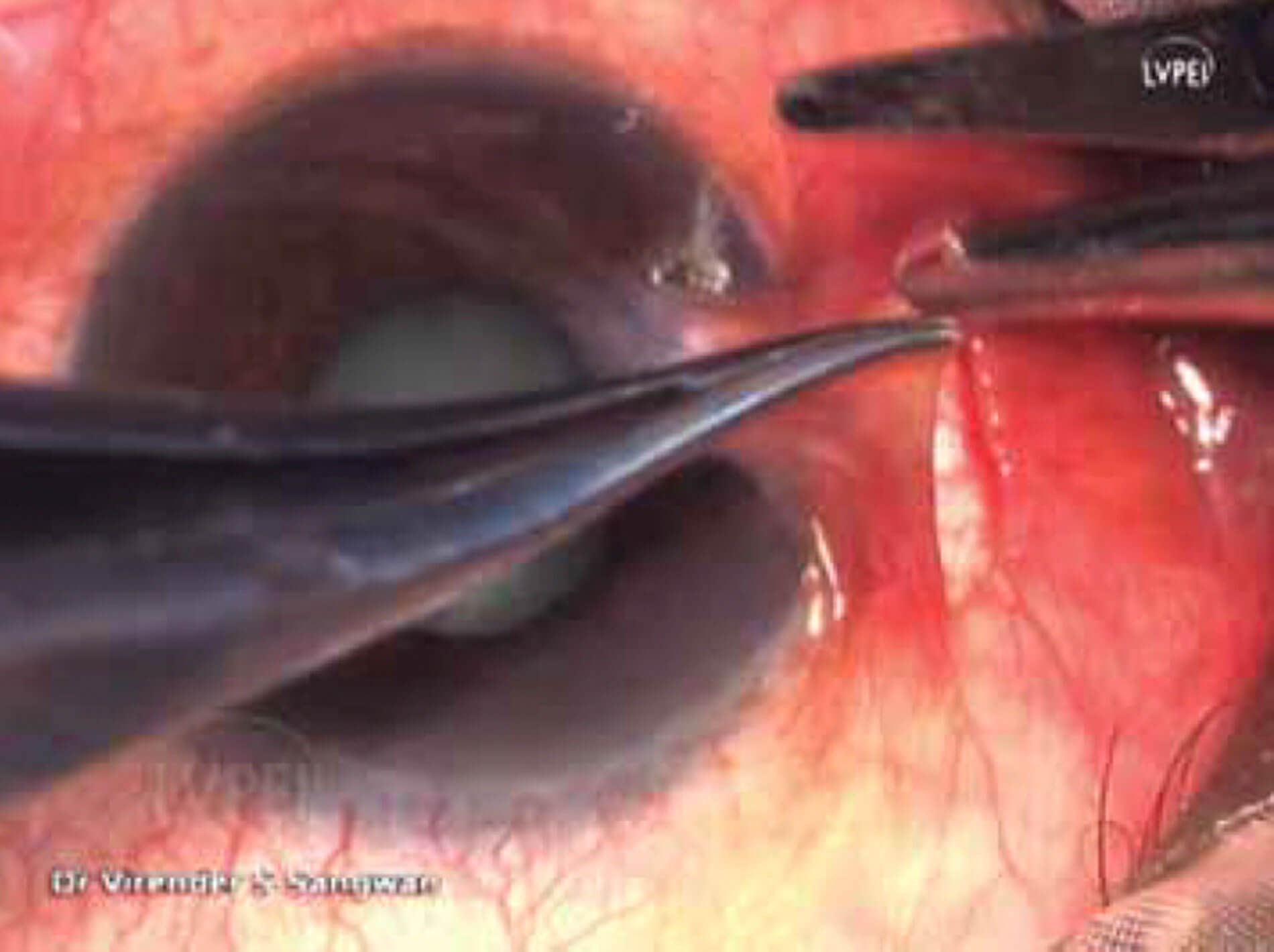
- Your physician will sedate you and administer local anesthesia to the eye to prevent pain. The surrounding area will be cleaned.
- The pterygium and a portion of the adjacent conjunctival tissue will be excised.
- After removal, the doctor will place a graft of conjunctival tissue or membrane to reduce the chance of the pterygium returning.
Sutures vs. glue
Following excision, the graft is secured either with sutures or with fibrin glue. Both approaches aim to lower the risk of recurrent pterygia.
Absorbable sutures are a longstanding method but can lead to greater postoperative discomfort and may prolong recovery for several weeks.
Fibrin glue, conversely, has been shown to lessen inflammation and pain and can shorten healing time (compared with sutures). Because fibrin glue is derived from blood products, it carries a small risk of transmitting viral infections or other diseases. It can also be more costly than suturing.
The bare sclera technique
An alternative is the bare sclera technique, which has a higher likelihood of recurrence. In this traditional method, the pterygium is removed but no graft is placed, leaving the exposed white of the eye to heal naturally.
While this technique avoids the potential complications related to sutures or fibrin glue, it is associated with a significant rate of pterygium regrowth, often resulting in larger lesions.
Recovery
At the conclusion of the operation, your doctor will typically apply a patch or pad over the eye for comfort and to reduce infection risk. Avoid rubbing the eye after surgery to prevent disturbing the graft.
Your physician will give specific postoperative care instructions, including cleaning routines, antibiotic use, and plans for follow-up appointments.
Complete healing — with reduction of redness and discomfort — can take from a few weeks to a few months, depending in part on the surgical technique used.
Complications
As with all surgeries, there are potential risks. Mild pain and redness are common after pterygium surgery. Temporary blurring of vision during recovery is also frequently reported.
If you experience worsening vision, sudden vision loss, or notice the pterygium returning, contact your eye care provider promptly.
Outlook
Pterygium surgery is generally successful. For mild cases, doctors may initially recommend topical medications and ointments. If these benign growths begin to impact vision or quality of life, surgical removal is usually the next option.
Frequently Asked Questions
Pterygium surgery involves excising the conjunctival growth, often followed by placement of a conjunctival graft or membrane to reduce recurrence. The procedure is usually done under local anesthesia with mild sedation.
Most pterygium surgeries last about 30 to 45 minutes. Time can vary slightly depending on whether sutures or fibrin glue are used and the complexity of the case.
Sutures are a traditional method but may cause more discomfort and longer healing. Fibrin glue often reduces inflammation and speeds recovery but is derived from blood products and can be more expensive.
Expect redness, mild pain, and temporary blurring as the eye heals over weeks to months. You will receive postoperative care instructions, including cleaning, medications, and follow-up visits.
Recurrence is possible, especially with the bare sclera technique. Using a conjunctival graft or membrane reduces the risk of regrowth, but follow-up care is important to monitor for recurrence.
Read Next
8 Ankle Stretches to Try at Home

Why Are Blisters Forming Between My Toes and How Do I Treat Them?

What Is Squid Ink, and Should You Eat It?
Proteolytic Enzymes: How They Work, Benefits and Sources
What Should I Do About a Molar Broken Off at the Gum Line

Multiple Sclerosis vs. Fibromyalgia: Differences in Signs and Symptoms
Related Posts

Binocular Vision Dysfunction: What You Should Know
Aug 21, 2026 • 15 min read
Dacryocystitis
Aug 25, 2026 • 14 min read
How to Use a Warm Eye Compress to Treat Styes and Other Conditions
Sep 9, 2026 • 28 min read
How Long Does It Take for the Effects from Eye Dilation Drops to Wear Off?
Jul 29, 2026 • 11 min read
How Many Calories Are in an Egg?
Aug 26, 2026 • 14 min read