Atherosclerosis of the Aorta: Plaque in Your Body's Largest Artery

- Your aorta in 30 seconds
- How plaque builds up in the aorta
- Thoracic vs abdominal aorta: different problems
- Symptoms: usually none — until something breaks off or blocks a branch
- Brain — emboli from the aortic arch
- Legs — emboli or branch narrowing
- Gut and kidneys — branch arteries starved of blood
- Back and abdominal pain — the wall itself
- The three dangers
- 1. Embolism — clots and cholesterol crystals
- 2. Aneurysm — the wall stretches
- 3. Dissection and rupture — the emergencies
- How doctors find it and measure it
- What the numbers mean
- ICD-10 code I70.0
- Treatment: slow it, stabilize it, protect the organs downstream
- Lifestyle — the foundation
- Medicines
- Procedures — for specific dangers, not for the aorta in general
- Can it be reversed?
- Your scan report says “aortic atherosclerosis” — what to do next
- The bottom line
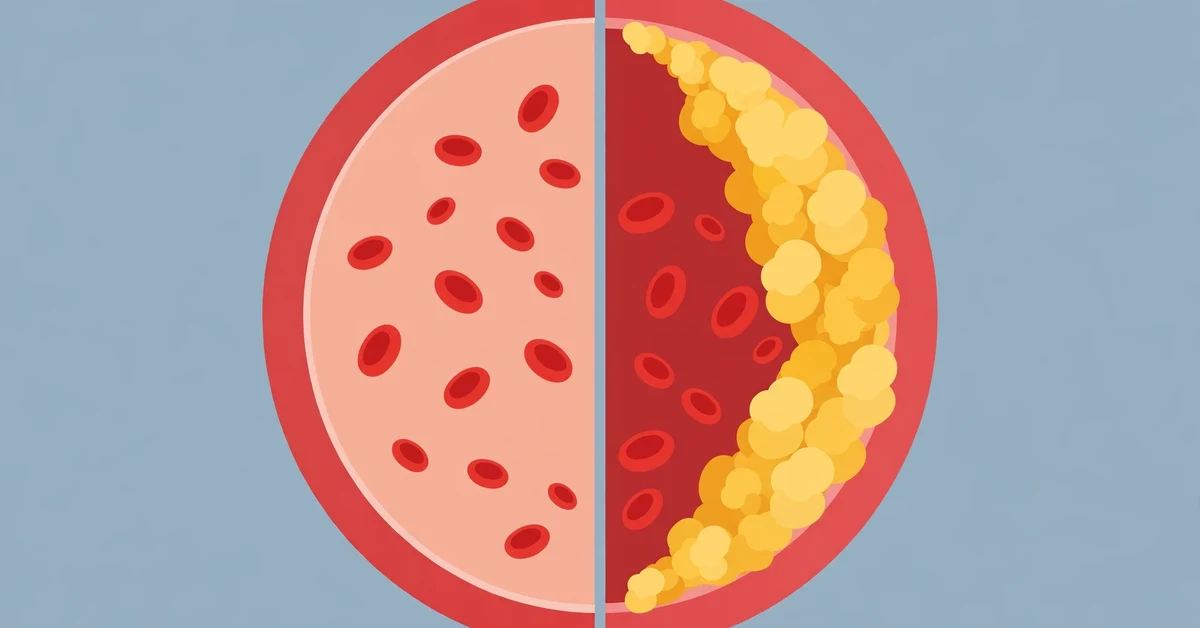
Atherosclerosis of the aorta is plaque buildup inside the wall of the aorta — the largest artery in your body. The aorta carries every drop of blood from your heart to the rest of your body, so plaque here matters more than plaque in a small artery. It usually causes no symptoms at all while it develops. Most people find out they have it by accident, when a chest X-ray or CT scan done for another reason reports “aortic calcification” or “atherosclerotic aorta.”
The danger is not the narrowing itself — the aorta is wide, about an inch across, and plaque rarely blocks it the way it blocks a heart artery. The danger is what the plaque can do: pieces of it, or blood clots that form on it, can break off and travel to the brain, kidneys, gut, or legs. And over years, plaque weakens the aortic wall, which can stretch into an aneurysm. Our overview of what atherosclerosis is explains the general process; this guide focuses on what happens when it strikes the aorta specifically.
Your aorta in 30 seconds
The aorta is the main highway of your circulation. It leaves the heart, curves over the top of the chest, runs down through the diaphragm, and ends in the abdomen, where it splits to supply the legs. Doctors divide it into four sections, because plaque in each section causes different problems:
- Ascending aorta — the short segment leaving the heart; it feeds the coronary arteries that supply the heart muscle itself.
- Aortic arch — the curve at the top of the chest; it gives off the arteries to the brain and arms.
- Descending thoracic aorta — runs down the back of the chest.
- Abdominal aorta — runs below the diaphragm and feeds the kidneys, the gut, and the legs.
Most aortic plaque is found in the abdominal aorta, below the diaphragm; a smaller share affects the chest (thoracic) aorta. The aorta is also where plaque most often calcifies — hardens with calcium deposits — which is why it shows up so clearly on X-rays and CT scans.

How plaque builds up in the aorta
The process is the same one described in our guide to when atherosclerosis starts, but the aorta’s size and flow patterns shape it in its own way. It begins when the inner lining of the artery (the endothelium) is injured — by high blood pressure, cigarette smoke, high blood sugar, or excess LDL cholesterol particles. Cholesterol slips into the wall and gets trapped there; immune cells move in to clean it up and become bloated “foam cells”; over years, dead cells, calcium, and scar tissue pile up into a plaque with a fibrous cap.
Two features of the aorta matter here. First, plaque forms most where blood flow is disturbed — at curves and branch points, such as the arch and the spot where the aorta splits in the abdomen. Second, aortic plaque calcifies heavily with age. A calcified plaque is stiffer and less likely to rupture than a soft, fatty one, but heavy calcification is a marker of a large total plaque burden — it tells doctors the disease is extensive, even if that particular plaque is stable.
Thoracic vs abdominal aorta: different problems
“Atherosclerosis of the aorta” is one diagnosis, but where the plaque sits changes what it can do to you.
| Thoracic aorta (chest) | Abdominal aorta (belly) | |
|---|---|---|
| Where the plaque is | Ascending aorta, arch, descending chest aorta | Below the diaphragm, often near the kidneys and the split to the legs |
| Main danger | Emboli travelling to the brain — stroke or TIA | Branch blockages (legs, gut, kidneys) and aneurysm |
| Typical symptoms | Usually none; sometimes chest or back pain if an aneurysm forms | Leg pain when walking, pain after eating, hard-to-control blood pressure; a pulsing lump if an aneurysm grows |
| How common | Smaller share of aortic plaque | Most aortic plaque is here |
The split is not absolute — many people have plaque in both sections, and a clot from the descending thoracic aorta can still reach the brain, because blood can flow backward in the arch under certain conditions. But as a rule of thumb: chest plaque threatens the brain, belly plaque threatens the legs, gut, kidneys — and the wall itself.
Symptoms: usually none — until something breaks off or blocks a branch
Atherosclerosis of the aorta is silent in most people. There is no pain, no warning, while the plaque grows. Symptoms begin when a piece of plaque or a clot breaks off and travels downstream, when a branch artery gets narrowed, or when the wall stretches into an aneurysm. Which symptoms appear depends on where the trouble lands:
Brain — emboli from the aortic arch
Sudden weakness or numbness on one side of the body, slurred or garbled speech, loss of vision in one eye, facial droop, or a sudden severe headache. These are the signs of a transient ischemic attack (TIA) or a stroke — a clot that broke off an aortic plaque and lodged in a brain artery. A TIA is a warning that a full stroke may follow, and it needs emergency assessment the same day.
Legs — emboli or branch narrowing
Cramping or aching in the calf, thigh, or buttock after walking a predictable distance, easing within minutes of rest — the same claudication described in peripheral artery disease. In more advanced disease: burning foot pain at rest, cool or pale feet, or sores on the feet that heal slowly.
Gut and kidneys — branch arteries starved of blood
Pain or cramping after meals, bloating, and unintentional weight loss from avoiding food can signal narrowed arteries to the intestines. Blood pressure that stays high despite several medications, or kidney function that slowly declines, can signal narrowed renal arteries.
Back and abdominal pain — the wall itself
A deep, steady ache in the back or abdomen can mean an aneurysm is growing. Sudden, severe pain in the same places is an emergency — see the box below. A pulsating lump felt in the abdomen is the classic sign of an abdominal aortic aneurysm, and it should always be checked promptly.

The three dangers
Aortic plaque harms people in three distinct ways. Understanding which one applies to you is the key to understanding your treatment.
1. Embolism — clots and cholesterol crystals
This is the most common danger, and it comes in two forms. Thromboembolism is the familiar one: a blood clot forms on the surface of a plaque, breaks off, and travels downstream until it wedges in a smaller artery — in the brain (stroke), the kidneys, the gut, or a leg. When tissue is starved of oxygen but not yet dead, doctors call it ischemia; once tissue dies, it is an infarction — we explain the difference in our guide to ischemia vs infarction.
Cholesterol embolization (atheroembolism) is the form almost no patient guide mentions. Here it is not a clot but tiny cholesterol crystals that shower downstream when a plaque cracks open — often triggered by a catheter procedure such as an angiography or angioplasty, or sometimes by starting a blood thinner. The crystals lodge in small vessels and cause a distinctive pattern: blue or purple toes, mottled purplish skin on the legs (livedo reticularis), and kidney function that falls over days to weeks. One clue that separates it from an ordinary blood clot is that the pulses in the feet are usually still felt — the blockage is in the tiny vessels, not the main leg artery. Diagnosis rests on the pattern, sometimes with a skin or kidney biopsy; treatment is supportive care, stopping the trigger, and protecting the kidneys.
2. Aneurysm — the wall stretches
Plaque and the inflammation around it damage the middle layer of the aortic wall (the media), which is what gives the aorta its strength and elasticity. A weakened patch can slowly bulge outward into an aneurysm. Most abdominal aortic aneurysms form below the kidney arteries, and most are tied to atherosclerosis. They usually grow silently, a few millimeters a year, and are often found by accident. The risk of rupture rises with size, and it rises sharply once an aneurysm passes about 5 centimeters (2 inches). Because of that, doctors watch small aneurysms with regular scans and usually start discussing repair around 5 to 5.5 centimeters, sooner if the aneurysm grows fast or causes pain.
3. Dissection and rupture — the emergencies
A dissection is a tear in the inner layer of the aortic wall; blood forces its way into the wall and splits it lengthwise. Atherosclerosis can weaken the wall and contribute, although dissection often strikes without any prior widening. A rupture is an aneurysm bursting. Both are life-threatening within minutes to hours, and both announce themselves with pain that is impossible to ignore.
Call 911 (or your local emergency number) immediately if you have:
- Stroke signs (BE-FAST): Balance loss, Eyes — sudden vision change, Face drooping, Arm weakness, Speech difficulty, Time to call 911.
- Sudden, severe, “tearing” chest or back pain — the classic sign of an aortic dissection. The pain often spreads and feels different from any chest pain you have had before.
- Sudden severe abdominal or back pain with dizziness, fainting, or clammy skin — possible rupture of an abdominal aortic aneurysm, especially if you can feel a pulsing lump in your belly.
Do not drive yourself. Note the exact time the symptoms started — it matters for treatment.
How doctors find it and measure it
Most aortic atherosclerosis is found by accident — a scan done for another reason mentions it. The aorta is hard to examine from the outside, and there is no routine screening test for aortic plaque itself. When doctors do look for it, they use these tools:
| Test | What it shows | When it’s used |
|---|---|---|
| Chest X-ray | Calcification outlining the aortic arch | Often the first, accidental finding |
| CT / CT angiography | Plaque, calcification, and aneurysm size in detail | The main test when an aneurysm is suspected or being measured |
| MRI | Plaque composition and wall detail, without radiation | When CT contrast is a problem, or for detailed wall assessment |
| Transesophageal echo (TEE) | Plaque in the arch and descending thoracic aorta, measured in millimeters | The best test for judging embolic risk from arch plaque |
| Abdominal ultrasound | Diameter of the abdominal aorta | Aneurysm screening and monitoring — cheap, no radiation |
| Blood tests | LDL, blood pressure, blood sugar, kidney function | They measure the drivers of plaque, not the plaque itself |
One common confusion: a coronary artery calcium (CAC) score measures plaque in the heart’s arteries, not the aorta. A high CAC score means you almost certainly have aortic plaque too, but the two scores are not interchangeable.
What the numbers mean
When a report describes aortic plaque, a few numbers decide how seriously doctors take it:
- Thickness of 2 mm or more on a transesophageal echo is the working definition of aortic atherosclerosis — the inner lining is thickened enough to count as plaque.
- Thickness of 4 mm or more in the aortic arch marks a “complex” plaque, especially if it is mobile or ulcerated. In a landmark study of stroke patients, plaques this thick in the arch were linked to a substantially higher risk of another stroke — which is why doctors treat arch plaque as a stroke risk, not just a finding.
- Calcification tells doctors about total plaque burden. Heavily calcified plaques are usually more stable than soft, fatty ones, but extensive calcification means the disease is widespread — the Multi-Ethnic Study of Atherosclerosis linked thoracic aortic calcification to higher mortality.
- Aneurysm diameter is the number that drives decisions: small aneurysms are watched with regular scans, and repair is usually discussed around 5 to 5.5 centimeters, or sooner if the aneurysm grows quickly or causes pain.
ICD-10 code I70.0
For reference: atherosclerosis of the aorta is coded I70.0 in ICD-10, the coding system used on medical records and insurance claims in the United States. It is a billable diagnosis code, valid for adults. If your medical record, hospital bill, or insurance paperwork lists I70.0, it means exactly this condition — plaque in the aorta. Related codes cover other arteries (for example, I70.1 for renal artery atherosclerosis), so the code also tells you which artery the diagnosis refers to.
Treatment: slow it, stabilize it, protect the organs downstream
There is no procedure that scrubs the aorta clean. Treatment has three goals: stop plaque from growing, stabilize the plaques that exist so they don’t throw emboli, and fix the specific danger — a branch narrowing or an aneurysm — when one appears.
Lifestyle — the foundation
- Quit smoking. The single highest-yield step. Smoke chemicals injure the artery lining directly and make blood more likely to clot; risk starts falling within weeks to months of quitting.
- Eat a Mediterranean-style pattern: vegetables, fruit, whole grains, legumes, nuts, olive oil, and fish; go easy on saturated fat, processed meat, sodium, and added sugar. See where your numbers fall in our cholesterol levels chart by age.
- Move most days: at least 150 minutes a week of moderate activity such as brisk walking.
- Control blood pressure and blood sugar — check your targets against our blood pressure chart by age, and treat sleep apnea if you have it.
Medicines
- Statins lower LDL cholesterol and stabilize plaque; for people with established atherosclerotic disease they are the backbone of treatment. For people at high risk, guidelines commonly target LDL below 70 mg/dL.
- Blood pressure medicines treat one of the main drivers of artery injury; the usual target is below 130/80 mm Hg.
- Antiplatelet medicines (such as low-dose aspirin or clopidogrel) reduce clot formation on plaque surfaces in people who already have atherosclerotic disease. (For people without disease, the benefit–bleeding balance is different — don’t start aspirin on your own.)
- Anticoagulants (blood thinners) are used more selectively — typically when a clot has already embolized, or when a mobile clot is seen sitting on an aortic plaque.
- Diabetes medicines treat high blood sugar, another driver of artery injury.
Procedures — for specific dangers, not for the aorta in general
- Endovascular aneurysm repair (EVAR) or open surgery for an abdominal aortic aneurysm that has reached repair size — a stent-graft placed inside the aorta to take pressure off the weakened wall.
- Thoracic endovascular repair (TEVAR) for a thoracic aneurysm or, in emergencies, for a dissection.
- Angioplasty and stenting, or bypass surgery, for a branch artery that is badly narrowed — for example, a kidney artery causing uncontrollable blood pressure, or leg arteries causing severe claudication.
These procedures fix a specific danger; they don’t change the disease in the rest of the artery tree, which is why lifestyle and medicines continue afterward.
Can it be reversed?
Honest answer: no — not completely, and not back to a brand-new aorta. The Cleveland Clinic states plainly that atherosclerosis of the aorta can’t be reversed, and that is the right expectation to carry. Calcified, scarred portions of old plaque do not melt away.
But the same nuance from our overview applies here: with intensive LDL lowering (high-intensity statins, sometimes combined with other drugs), imaging studies inside arteries have measured small but real shrinkage of soft plaque — on the order of about 1% of total atheroma volume over 1–2 years. More important than shrinkage is stabilization: treated plaques develop thicker caps and smaller fatty cores, making the rupture-and-embolus event — the actual cause of most strokes from aortic plaque — much less likely. Event rates fall far more than the small change in narrowing would suggest.
So the realistic goal is: stop the growth, stabilize what’s there, and prevent the event. People who quit smoking, get LDL and blood pressure to target, and stay active can live for decades with aortic atherosclerosis without a stroke or a ruptured aneurysm. The worst strategy is assuming a “mild” finding means no action is needed — mild plaque is exactly the stage where treatment works best.
Your scan report says “aortic atherosclerosis” — what to do next
If those words just appeared on your results, work through this list with your doctor:
- Don’t panic. Aortic plaque is common, often mild, and usually found by accident. The finding is a reason to act, not a verdict.
- Ask where and how bad: thoracic or abdominal? How thick is the plaque? Is there an aneurysm, and if so, how wide? Is any branch artery narrowed?
- Get the drivers measured: LDL cholesterol, blood pressure, blood sugar (A1C), and kidney function. Ask about lipoprotein(a) — a cholesterol-like particle set by your genes that is worth measuring once in a lifetime.
- If you smoke, quitting is the single most effective step — more than any single medicine.
- Ask whether you need further tests: a CT angiography to map the plaque, an abdominal ultrasound to check for an aneurysm, or a referral to cardiology or vascular surgery.
- Know the red flags: stroke signs (BE-FAST), sudden tearing chest or back pain, or sudden severe abdominal pain with dizziness — all mean calling 911, not waiting for an appointment.
- Set a monitoring plan: if there is an aneurysm, your doctor will set a scan interval based on its size; if there is none, the plan is usually risk-factor control plus re-imaging only if symptoms appear.
The bottom line
Atherosclerosis of the aorta is plaque in the body’s largest artery. It rarely blocks blood flow the way plaque in a heart artery does — instead, its dangers are emboli that travel to the brain, kidneys, gut, or legs, and a weakened wall that can stretch into an aneurysm. It is usually silent and usually discovered by accident on a scan, which is why a report mentioning “aortic calcification” deserves attention rather than dismissal.
The hopeful part: its main drivers are measurable and treatable. Know your LDL, blood pressure, and blood sugar; don’t smoke; keep moving; and if a scan has already shown aortic plaque, ask your doctor where it is, how severe it is, and whether there is an aneurysm to watch. If warning signs of a stroke or a ruptured aneurysm appear, treat them as the emergency they are and call 911.
This article is general information only and is not medical advice. If you are concerned about your aorta or your cardiovascular risk, talk to your own doctor, who can interpret tests in the context of your health history.
Sources: Cleveland Clinic, What is Atherosclerosis of the Aorta? ; National Heart, Lung, and Blood Institute (NHLBI), What Is Atherosclerosis? ; American Heart Association, What Is Atherosclerosis?; Mayo Clinic, Arteriosclerosis/Atherosclerosis — Symptoms and Causes ; Amarenco et al., 1994, on aortic arch plaque thickness and stroke risk; U.S. Preventive Services Task Force, abdominal aortic aneurysm screening recommendation.
Frequently Asked Questions
Atherosclerosis of the aorta is the buildup of fatty plaque inside the wall of the aorta, the largest artery in the body. The plaque narrows and stiffens the vessel, but the main dangers are different from a blocked heart artery: pieces of plaque or blood clots can break off and travel to the brain, kidneys, gut, or legs, and the weakened wall can stretch into an aneurysm.
Usually there are none — early aortic atherosclerosis is silent. Symptoms appear when an embolus travels downstream (stroke or TIA signs, leg cramps when walking, pain after eating, hard-to-control blood pressure) or when an aneurysm grows (deep back or abdominal pain, a pulsating lump in the belly). Because symptoms start late, most cases are found by accident on a scan.
It can be. The serious risks are stroke from emboli breaking off arch plaque, kidney failure or leg ischemia from emboli, and rupture of an abdominal aortic aneurysm. But many people live for decades with mild aortic plaque that is controlled with statins, blood pressure treatment, and not smoking. The danger depends on the plaque’s location, thickness, and whether an aneurysm has formed.
I70.0. It is the billable ICD-10 diagnosis code for atherosclerosis of the aorta, used on medical records and insurance claims in the United States. If your paperwork lists I70.0, it means plaque in the aorta specifically.
Most often it is found incidentally — a chest X-ray or CT scan done for another reason reports aortic calcification. When doctors look for it deliberately, they use CT or CT angiography to map plaque and aneurysms, MRI for wall detail, transesophageal echo (TEE) to measure arch plaque thickness, and abdominal ultrasound to measure the abdominal aorta. Blood tests measure the drivers (LDL, blood pressure, blood sugar), not the plaque itself.
No, not completely — calcified plaque does not dissolve, and the Cleveland Clinic states plainly that it can’t be reversed. But intensive treatment can stabilize plaque and slightly shrink its soft parts: high-intensity statins (sometimes combined with other drugs), not smoking, and blood pressure control make plaques less likely to rupture and throw emboli, which is what actually prevents strokes and other events.
Thoracic aortic atherosclerosis affects the chest portion of the aorta and mainly threatens the brain — emboli from arch plaque cause strokes. Abdominal aortic atherosclerosis affects the belly portion, is more common, and mainly threatens the legs, gut, and kidneys through branch narrowing, plus the wall itself through aneurysm. Many people have plaque in both sections.
Don’t panic — it is common and often mild. Ask your doctor where the plaque is (thoracic or abdominal), how severe it is, and whether there is an aneurysm. Get your LDL, blood pressure, blood sugar, and kidney function checked; quit smoking if you smoke; and ask whether you need a CT angiography, an abdominal ultrasound, or a cardiology referral. Learn the red flags — stroke signs and sudden severe chest, back, or abdominal pain — and call 911 if they appear.
Read Next
Does Medicare Cover CPAP Machines? Coverage Rules and Costs Explained

How to Do Chair Dips
Salmonella Food Poisoning

9 Stretches for Sciatica Pain Relief
Bedsore Stages in Pictures: What Each Pressure Ulcer Stage Looks Like
Blood Pressure Chart by Age: Average Readings, Healthy Targets and What Your Numbers Mean
Related Posts
What Is Atherosclerosis? Plaque Buildup in Your Arteries, Explained
Nov 11, 2026 • 17 min read
What Causes Peau D’Orange?
Aug 23, 2026 • 20 min read
9 of the Best Keto-Friendly Protein Bars You Can Buy
Sep 17, 2026 • 31 min read
Side Effects of Brixadi: What You Need to Know
Aug 28, 2026 • 11 min read
Hernias by Picture
Sep 26, 2026 • 22 min read