What Are Back Mice and How Are They Treated?


Back mice is an informal phrase used to describe painful nodules located on or near the lower back and hip regions.
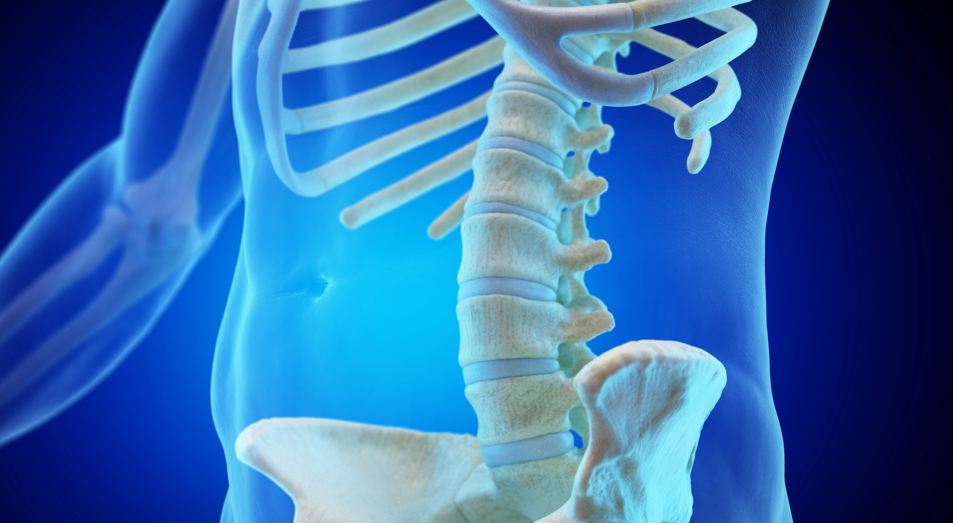
A 2016 article in Pain Physician characterizes back mice as “firm, rubbery, mobile nodules that are located in characteristic regions of the sacroiliac, posterior superior iliac, and the lumbar paraspinal regions.”

The label “back mice” was first introduced by Peter Curtis in 1993 to describe episacral (or episacroiliac) lipomas. Today the term can apply to several different diagnoses.
Keep reading to learn more about what back mice might indicate and available treatment approaches.
Conditions often referred to as back mice
Frequently labeled as nonspecific low back pain, back mice is a lay term that could denote multiple conditions, including:
- episacral lipoma
- iliac crest pain syndrome
- lumbar fascial fat herniation
- lipoma
Episacral lipoma
An episacral lipoma is a small, tender nodule located beneath the skin. These painful bumps develop when a portion of the dorsal fat pad pushes through a defect in the thoracodorsal fascia (the connective tissue that supports the back muscles).
They most commonly appear over the posterior iliac crest — the upper, outer border of the pelvic bone.
Iliac crest pain syndrome
Iliac crest pain syndrome, sometimes called iliolumbar syndrome, stems from a tear in the iliolumbar ligament. Repeated bending or twisting movements can cause this ligament to become damaged.
It may also follow trauma such as a fall, sports-related injury, or motor vehicle collision.
Lumbar fascial fat herniation
Lumbar fascial fat herniation refers to a painful fatty mass that protrudes through the lumbodorsal fascia.
The lumbodorsal fascia is a thin, fibrous layer that overlays the deep muscles of the back.
Lipoma
A lipoma is an overgrowth of fat cells forming a lump beneath the skin. Lipomas are benign (noncancerous) tumors.
When a fatty lump is detected in the lower back area, the immediate assumption is often a lipoma, though some people may call it a back mouse.
Other problems sometimes called back mice
Additional conditions that are occasionally labeled as back mice include:
- Sebaceous cyst. A fluid-filled sac that forms within the skin layers (between the epidermis and dermis). Like lipomas, sebaceous cysts are benign.
- Subcutaneous abscess. In this situation, pus accumulates beneath the skin. Subcutaneous abscesses are typically painful and later become swollen and red.
- Liposarcoma. These are malignant tumors that can present as fatty masses within muscle tissue.
Typical treatments for back mice
At present, management of back mice most commonly involves local injections of an anesthetic such as lidocaine, often combined with corticosteroids.
A 2000 study reported that 86 percent of participants were “satisfied” or “very satisfied” after receiving injections of local anesthetic and corticosteroid.
Although injections relieved both local pain and radiating symptoms, they did not always remove the palpable nodules. The study reported no adverse events.
Takeaway
Back mice (or a back mouse) is an informal term for painful lumps on the lower back or hips. Because it’s not a formal medical diagnosis, the term may be used to describe several different conditions, such as:
- episacral lipoma
- subcutaneous abscess
- liposarcoma
- iliac crest pain syndrome
- lumbar fascial fat herniation
If you notice painful bumps on your hips or lower back, consult a healthcare professional. They can determine the underlying cause and suggest an appropriate treatment plan.
Frequently Asked Questions
Back mice is an informal term for small, often painful nodules or fatty lumps that appear near the lower back and hip regions; they can represent conditions like episacral lipoma, fascial fat herniation, or other subcutaneous masses.
Symptoms commonly include localized tenderness or sharp pain at the site of the nodule, sometimes with radiating discomfort; visible or palpable bumps may be present, and symptoms can worsen with movement or pressure.
Diagnosis typically starts with a physical exam and palpation of the tender nodule; imaging (ultrasound, MRI) or referral to a specialist may be used to differentiate lipoma, abscess, herniation, or other causes.
Treatment often includes local injections of anesthetic and corticosteroid to reduce pain and inflammation; other options depend on the underlying cause and can include surgical removal or management of infection when indicated.
Consult a healthcare professional if you have a painful lump, worsening pain, signs of infection (redness, warmth, fever), or any rapidly growing mass to obtain an accurate diagnosis and appropriate treatment plan.
Read Next

Midlife Crisis or Midlife Myth? What to Know About Going ‘Over the Hill’
The 5 Best Thumb Braces for Arthritis

5 Evidence-Based Ways Collagen May Improve Your Hair
7 Ways to Remove Dead Skin from Your Feet

Can You Use Face Mapping to Improve Your Skin’s Health?
Natural Treatment for Seborrheic Dermatitis: What Works?
Related Posts

Stretches and Exercises to Help Ease SI (Sacroiliac) Joint Pain
Oct 5, 2026 • 36 min read
Pregnancy Rib Pain: Causes, Prevention, Remedies
Sep 19, 2026 • 23 min read
6 Foam Roller Exercises for Your Back
Sep 16, 2026 • 36 min read
12 Spinal Decompression Exercises for All Skill Levels
Sep 21, 2026 • 12 min read
10 Back Exercises with Bands to Counteract Work-from-Home Posture
Sep 14, 2026 • 21 min read