Ultrasounds Aren't Typically Used to Detect Cervical Cancer: Learn Why

- Can you see cervical cancer on an ultrasound?
- What tests can detect cervical cancer?
- Pap tests for cervical cancer
- HPV test for cervical cancer
- Colposcopy
- What tests can diagnose cervical cancer?
- Imaging tests for people with cervical cancer
- Types of ultrasound
- Abdominal ultrasound
- Pelvic and transvaginal ultrasound
- How often should you be screened for cervical cancer?
- Takeaway
Ultrasounds are not commonly used to identify cervical cancer and should not replace routine screening methods. There are simpler, more dependable approaches for screening and diagnosing cervical cancer.
An ultrasound is not a primary tool for detecting cervical cancer because superior screening options exist. During routine gynecologic visits, clinicians typically screen for cervical cancer using a Pap smear and a human papillomavirus (HPV) test. These exams assess risk factors and search for abnormal cell changes on the cervix.
Cervical cancer is among the most preventable cancers due to the ease of screening. If a clinician finds suspicious findings, they may order imaging tests, but ultrasound is not usually the first choice.
Read on to understand the role of ultrasound in cervical cancer and to learn about more effective screening and diagnostic procedures.
Can you see cervical cancer on an ultrasound?
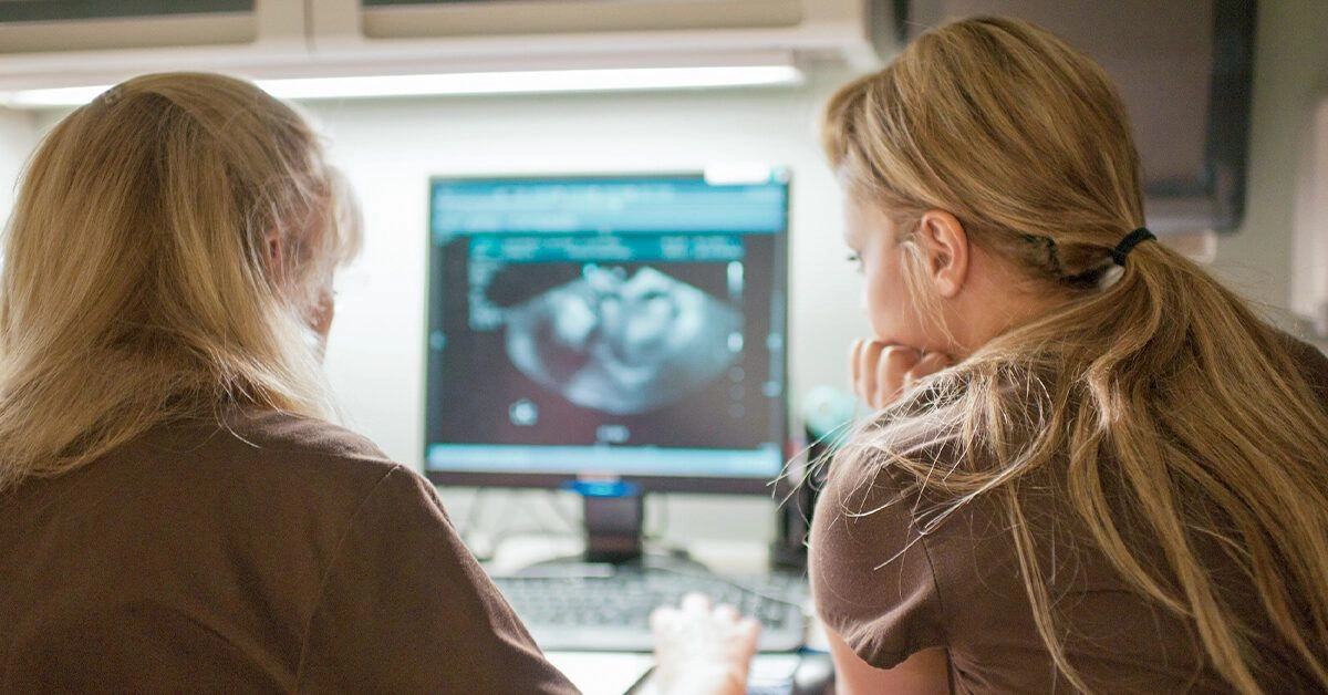
An ultrasound is not the ideal method to screen for or confirm cervical cancer. Ultrasound uses sound waves to produce images of internal structures and is frequently employed in obstetrics and gynecology to follow fetal growth and to identify fibroids, cysts, and other masses.
Although certain ultrasound techniques can reveal signs of some cervical cancers, clinicians rarely spot cervical cancer incidentally during an exam aimed at another issue. The sonographer performing the study may not be trained to detect cervical cancer, particularly if the scan is being done for a different reason.
More accurate ways to screen for and diagnose cervical cancer include:
- Pap smear
- HPV testing
- Colposcopy
- Biopsy
That said, ultrasound can be part of the evaluation later in the diagnostic process. After a biopsy confirms cervical cancer, a physician may request various imaging studies to determine whether the disease has extended into adjacent tissues or metastasized elsewhere.
What tests can detect cervical cancer?
Screening for cervical cancer generally involves a Pap test, an HPV test, or a combination of both.
Routine screening is crucial for catching this cancer early when it’s most treatable. It’s estimated that about half of cervical cancer cases occur in people who have never had Pap or HPV testing.
Pap tests for cervical cancer
A Pap test checks for abnormal cells on the cervix and is usually performed during a standard gynecological exam. A clinician inserts a speculum into the vagina and uses a brush or spatula to collect a cellular sample, which is then analyzed in a laboratory.
If your Pap result is “abnormal,” further evaluation may be advised to confirm or exclude cervical cancer. Your provider might also schedule closer surveillance with more frequent exams.
HPV test for cervical cancer
The most frequent cause of cervical cancer is infection with HPV. Performed during a pelvic exam, HPV testing checks for this virus. The advantage is that clinicians can detect HPV before it causes precancerous alterations in cervical cells.
HPV is very common, and most infections do not progress to cancer. There are over 100 HPV strains, and some are linked to a higher cervical cancer risk.
HPV testing can be particularly helpful if you haven’t received the HPV vaccine or haven’t completed the vaccination series. Nevertheless, everyone with a cervix should continue regular Pap testing because the current vaccine does not protect against all HPV strains.
Colposcopy
Pap and HPV tests serve as screening tools. When these results are abnormal, clinicians usually recommend additional evaluation, typically starting with a colposcopy. This procedure uses a magnifying instrument called a colposcope to examine the cervix’s surface closely.
This exam is similar to a pelvic exam. A speculum is inserted into the vagina and the clinician inspects the cervix through the colposcope. They might apply a mild acidic solution to highlight abnormal areas, which generally does not add discomfort.
What tests can diagnose cervical cancer?
If colposcopy reveals suspicious areas, a doctor will advise a biopsy, which involves taking a small tissue sample for laboratory analysis. This can be performed during the colposcopy or at a later appointment.
Biopsy methods used to diagnose cervical cancer include:
- Colposcopic biopsy: Taken during a colposcopy, the clinician removes a small piece of tissue from the identified abnormal area.
- Cone biopsy (conization): A cone-shaped portion of cervical tissue is excised. This approach is also therapeutic for removing early-stage cervical cancer.
- Endocervical scraping (endocervical curettage): Often used when other biopsies are inconclusive, this extracts tissue from the endocervical canal.
If a biopsy is positive for cancer, physicians may order more tests to determine the extent of spread, which can include:
- A pelvic exam
- A rectal exam
- Cystoscopy to inspect the bladder and urethra
- Colonoscopy to examine the colon
Imaging tests for people with cervical cancer
If there is concern that cervical cancer may have spread, imaging studies can help confirm the suspicion and guide treatment planning. These studies also help determine the cancer’s stage.
Imaging may include the following:
- Chest X-rays to check for metastasis to the lungs.
- Intravenous (IV) urography, a type of X-ray that evaluates the urinary tract, including kidneys.
- Computed tomography (CT) scans of the pelvis or other areas to measure tumor size and look for spread.
- Combined CT and positron emission tomography (PET) scans for more detailed images.
- Magnetic resonance imaging (MRI), which can be superior to CT for visualizing soft tissues.
Types of ultrasound
There are multiple ultrasound modalities, each serving to visualize different internal regions and conditions.
Abdominal ultrasound
An abdominal ultrasound images abdominal organs like the liver, spleen, and pancreas, and can assess blood flow in these structures. Physicians may request an abdominal ultrasound to evaluate:
- blood clots
- accumulation of fluid or pus
- infections
- obstructions
- cysts or tumors
- fetal development during pregnancy
However, abdominal ultrasound is not intended for detailed imaging of the cervix or the broader pelvic area.
Pelvic and transvaginal ultrasound
A pelvic ultrasound images tissues, organs, and structures within the pelvis, such as the cervix, uterus, fallopian tubes, and vagina.
Pelvic ultrasound is performed externally on the abdomen, whereas a transvaginal ultrasound uses a probe introduced into the vagina for closer images.
While pelvic ultrasound can reveal fibroids and other pelvic masses, it is not the preferred diagnostic tool for cervical cancer.
Physicians may order transabdominal or transvaginal pelvic ultrasound to evaluate:
- possible structural irregularities
- pelvic inflammation or infections
- fetal growth during pregnancy
- suspected ectopic pregnancy
- potential fetal abnormalities
- blood flow in pelvic organs
- the cervix’s overall thickness and length
How often should you be screened for cervical cancer?
Regular screening is the most effective way to detect cervical cancer early, before it spreads. The American Cancer Society reports that the 5-year relative survival rate for localized cervical cancer is 92%.
Accordingly, the Centers for Disease Control and Prevention (CDC) recommends the following screening schedule:
| Ages | Screening Recommendations |
|---|---|
| Age 21 | Begin Pap testing. |
| Ages 21 to 29 | If Pap results are normal, repeat every 3 years. |
| Ages 30 to 65 | Options include: Pap test only. If normal, every 3 years. HPV test only. If negative, every 5 years. Pap and HPV co-testing. If both normal, every 5 years. |
| Age 65+ | If recent results have been consistently normal, screening can be stopped. |
Remember to report any unusual changes between scheduled screenings. Speak with a clinician if you experience symptoms such as:
- abnormal spotting or bleeding
- very heavy menstrual bleeding
- unusual vaginal discharge
- pelvic pain
- bleeding or pain after intercourse
Takeaway
While you may have an ultrasound for other medical reasons, for example during pregnancy, it should not replace routine cervical cancer screening. Staying current with recommended tests helps identify problems early.
If abnormal cell changes are detected on a Pap test, or if an HPV test is positive, your clinician will recommend appropriate follow-up. Contact your healthcare provider if you notice any concerning symptoms — earlier detection generally leads to better outcomes.
Frequently Asked Questions
An ultrasound can sometimes show irregularities in pelvic tissues, but it is not a reliable screening or diagnostic tool for cervical cancer. Pap smears, HPV testing, colposcopy, and biopsy are the preferred methods for detecting and confirming cervical cancer.
Ultrasound may be used after a biopsy confirms cervical cancer to assess tumor size or spread to nearby pelvic structures, but it is typically not the first-line imaging choice for staging; MRI, CT, or PET/CT are often preferred.
Routine screening is done with a Pap smear and/or an HPV test. These tests detect abnormal cervical cells or high-risk HPV strains before cancer develops, allowing for early intervention.
No. Ultrasound can detect masses like fibroids or cysts that are usually benign. A suspicious finding requires further evaluation with colposcopy and biopsy to determine if cancer is present.
Screening recommendations vary by age: begin Pap testing at 21; ages 21–29 repeat Pap every 3 years if normal; ages 30–65 options include Pap every 3 years, HPV test every 5 years, or co-testing every 5 years. Follow your clinician’s guidance.
Read Next

Arbonne Diet Review: Overview, Effectiveness, and More

Karate vs. Taekwondo: What's the Difference?

The 10 Best Selenium Supplements, According to a Dietitian

Incubators for Babies: Why They’re Used and How They Work

White Mushrooms: Nutrition, Benefits, and Uses

Carob Powder: 9 Nutrition Facts and Health Benefits
Related Posts

Is the Pain in My Tailbone Cancer?
Aug 21, 2026 • 15 min read
Low Sodium Frozen Meals: The Best Brands and 21 Dietitian-Approved Options
Sep 15, 2026 • 28 min read
How to Have Next-Level Car Sex
Sep 25, 2026 • 22 min read
Does Flaxseed Really Work for Beautiful Hair?
Aug 4, 2026 • 22 min read
Yoga for Endometriosis: Try These Stretches to Relieve Pain
Sep 18, 2026 • 4 min read