Hidradenitis Suppurativa Pictures: What Each Hurley Stage Looks Like

- Hidradenitis suppurativa at a glance: the signs to look for in photos
- The Hurley stages at a glance
- Stage 1 (mild) hidradenitis suppurativa pictures
- Stage 2 (moderate) hidradenitis suppurativa pictures
- Stage 3 (severe) hidradenitis suppurativa pictures
- Hidradenitis suppurativa by body area
- What hidradenitis suppurativa looks like on different skin tones
- Is it HS or something else? Look-alikes, side by side
- A 3-question self-check — and how doctors confirm HS
- Flares, and the warning signs that need prompt care
- What your stage means for treatment
- How to take photos of HS that actually help your doctor
- The bottom line
Hidradenitis suppurativa (HS), also called acne inversa, is a long-term inflammatory skin disease that begins in hair follicles where skin rubs against skin — the armpits, groin, buttocks and under the breasts. It produces deep, painful lumps that can break open, drain, heal and then come back in the same place. Because early HS is so often mistaken for ordinary boils, acne or ingrown hairs, people wait an average of about seven years for the correct diagnosis. Learning what it actually looks like is the fastest way to shorten that wait.
This page is a photo guide organised by the three Hurley stages dermatologists use to grade HS severity. Every photo is captioned with what to look for, and later sections show how HS differs from the conditions it is confused with. Two things first: HS is not contagious and is not caused by poor hygiene — it is an inflammatory disease of the hair follicle, as the Mayo Clinic and the American Academy of Dermatology emphasise. And the stage 3 photographs are graphic, because stage 3 itself is: they are included so anyone at that stage recognises it and gets specialist care.
Hidradenitis suppurativa at a glance: the signs to look for in photos
Whatever the stage, HS pictures share a recognisable pattern. Look for these six features — a single boil has one or two of them; HS has most of them together:
- Location in skin folds. Armpits, groin creases, inner thighs, buttocks, the fold under the breasts, waistband lines. HS almost never starts on flat, dry skin like the shin or forearm.
- Deep, firm, painful lumps. The bumps sit deep under the skin like marbles, not on the surface like pimples, and they hurt out of proportion to their size.
- Blackheads in pairs. Small pitted areas of skin dotted with blackheads, often two joined together (“double-ended” or tombstone comedones) — noted by DermNet NZ and the Mayo Clinic.
- Recurrence in the same spots. A lump heals, then weeks or months later another rises in almost the same place.
- Tunnels under the skin. In later stages, lumps connect beneath the surface, forming channels (sinus tracts) that drain through small openings.
- Rope-like scars. Thick, banded or bridging scars across the fold — the long-term footprint of repeated flares.
The Hurley stages at a glance
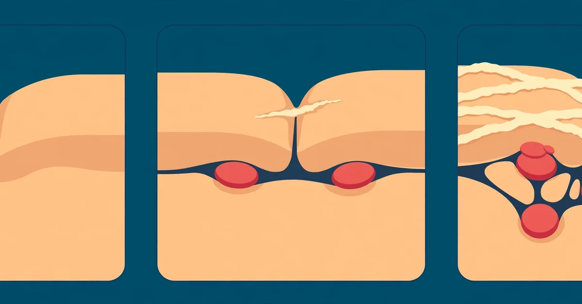
The Hurley system, introduced in 1989, grades HS by how much permanent change — tunnels and scarring — has built up in an area. A person can be stage 1 in the groin and stage 3 in one armpit at the same time — each area is staged separately.
| Hurley stage | Abscesses | Tunnels (sinus tracts) | Scarring |
|---|---|---|---|
| Stage 1 (mild) | One or more, isolated from each other | None | None |
| Stage 2 (moderate) | Recurring, widely separated in the same region | Present | Present |
| Stage 3 (severe) | Many, merged across a whole area | Multiple, interconnected | Extensive |
Hurley staging maps accumulated damage, not how active the disease feels this month — a stage 2 area can sit quiet for weeks, then flare hard. Doctors therefore also count inflamed nodules and draining tunnels when judging treatment response.
Stage 1 (mild) hidradenitis suppurativa pictures
Stage 1 HS looks deceptively ordinary. It begins with one or a few tender, pea-sized lumps deep in the skin of a fold — an armpit, the groin crease, under a breast. The skin over the lump may look pink, red, purple or just swollen, and itching, burning or extra sweating in the spot often comes a day or two before the lump itself. Small blackheads, often in pairs, may dot the surrounding skin. There are no tunnels and no scars yet.
Because a stage 1 lump resembles a boil, an ingrown hair or a deep pimple, it is usually treated as one — until it keeps coming back. A stage 1 lump typically stays sore for one to several weeks. It may soften, leak a little fluid and slowly settle, or shrink without ever opening. Weeks or months later a new one appears in the same fold — and that recurrence is the single most important clue in these photographs.


What to look for in stage 1 photos: lumps that sit deep rather than on the surface; normal, unscarred skin between lumps; blackhead pairs nearby; and — invisible in a single photo, but it decides the diagnosis — the same fold producing lumps again and again.
Stage 2 (moderate) hidradenitis suppurativa pictures
In stage 2 the pattern changes in two visible ways. The lumps keep returning and no longer fully clear between flares, and healing starts to leave marks: the first flat or pitted scars appear between lesions, and narrow channels — sinus tracts, often simply called tunnels — begin connecting lumps beneath the skin. On the surface, a tunnel may show only as a small opening that leaks pus or blood-stained fluid on and off for months, or as a firm cord you can feel running under the skin.
Stage 2 lesions are still widely separated — there is normal skin between them — and that separation is exactly what stage 3 loses. Flares at this stage are more painful, drain more, and the drained fluid often has the distinctive unpleasant smell people with HS know well. The pus itself does not mean the skin is infected; it is produced by the inflammation, although a secondary infection can develop on top.


What to look for in stage 2 photos: scars sitting between active lumps; small openings that never quite close; cord-like lines under the skin; and lesions dotted across one region rather than in a single spot.
Stage 3 (severe) hidradenitis suppurativa pictures
Stage 3 is HS that has taken over an entire area. The lumps merge into broad, swollen plaques; the tunnels underneath connect into a network, so pressing one place can make fluid appear at an opening some distance away. Several openings drain pus and blood more or less constantly, with the odour that causes so much distress. Thick, rope-like scars cross the whole fold and can physically restrict movement — raising an arm or walking becomes painful.
Scar tissue can also block lymph drainage, so long-standing stage 3 may cause persistent swelling of a limb or the genitals. Stage 3 is the end point of years of undertreated stage 1 and 2 disease — which is why the earlier sections of this page matter more than this one.


What to look for in stage 3 photos: no healthy skin between lesions; multiple draining openings in one connected field; discoloured, thickened plaques; and wide bands of scar tissue.
Hidradenitis suppurativa by body area
The stage pictures above follow the same rules everywhere, but each site has its own habits — and its own look-alikes.
| Body area | What HS tends to look like there |
|---|---|
| Armpits | The most photographed site: deep nodules in the vault of the armpit, later tunnels and scars that can limit arm movement. |
| Groin and inner thighs | Lumps along the crease and inner thigh; in women, lesions can also appear on the vulva and be mistaken for Bartholin cysts. |
| Under the breasts | Nodules and tunnels in the fold beneath the breast; see our dedicated guide to hidradenitis suppurativa on the breast for site-specific pictures and care. |
| Buttocks and around the anus | Deep nodules across the buttocks; around the anus, HS can mimic or accompany fistulas and needs specialist assessment. |
| Waist and belly folds | Lesions track along the waistband line and under an abdominal fold, as in the stage 3 abdomen photo above. |
| Less common sites | The nape of the neck, behind the ears and under a belt line. HS behind the ear is uncommon enough to have its own pattern — covered in our guide to hidradenitis suppurativa of the ear. |
Notice what is missing from that list: flat, exposed skin. A spreading flat rash on the lower legs, for example, is almost never HS — compare pictures of skin rashes on the lower legs to see the difference.
What hidradenitis suppurativa looks like on different skin tones
Most HS photographs online — including several above — show lighter skin, where inflammation reads as pink or red. On medium and deep skin tones the same disease looks different — one reason HS is under-recognised in people of colour. Active nodules may appear purple, dark brown or grey rather than red, and the redness around a flare may be barely visible; the swelling, tenderness and firmness you can feel are more reliable than colour. Between flares, the dominant marks are often dark patches (post-inflammatory hyperpigmentation) and rope-like scars.
So do not rule HS out because your lumps do not look “red enough” compared with photos. The deciding features — deep painful lumps in folds that keep returning and eventually tunnel and scar — are the same on every skin tone.
Is it HS or something else? Look-alikes, side by side
Several common conditions look very similar to HS in a single photograph. What separates them is the pattern over time and a few specific details.
| Condition | What it looks like | The difference that gives it away |
|---|---|---|
| Boil (furuncle) | One hot, red, pus-filled lump, often with a central pustule | Acute and one-off: it comes to a head, drains and is gone within about two weeks, leaving no tunnels. It does not keep returning to the same fold |
| Folliculitis | Many tiny surface pustules, each centred on a hair | Superficial and small — pinhead-sized spots on the skin surface, not deep marble-like lumps; no tunnels or rope-like scars |
| Ordinary skin abscess | A single tender, fluctuant pocket of pus, often after a cut or bite | Usually one episode with an obvious trigger and a bacterial cause; HS abscesses recur in chains in folds without any injury |
| Epidermal (sebaceous) cyst | A smooth, slow-growing dome under the skin | Typically has a tiny central punctum (pore) on its surface — a feature HS lumps do not have. Cysts are painless unless infected |
| Acne | Blackheads, whiteheads and pimples on the face, chest or back | Stays in acne territory (face, upper back), lesions are smaller and shallower, and it does not produce tunnels or draining openings in folds |
| Bartholin cyst | A swelling at one side of the vaginal opening | Comes from a single blocked gland in one specific spot; HS in the groin produces multiple lumps, blackhead pairs and lesions in other folds too |
The folliculitis row deserves a second look, because early HS and folliculitis are confused in both directions. Compare our folliculitis pictures: the pustules sit on the surface around hairs, while even the mildest HS lump in the photos above is clearly deep.
A 3-question self-check — and how doctors confirm HS
Dermatologists diagnose HS from three features, sometimes called the Dessau criteria. You can run the same check against your own photos:
- Typical lesions? Deep, painful nodules, abscesses, draining tunnels or rope-like scars — not just surface pimples.
- Typical places? Armpits, groin, buttocks, under the breasts or other skin folds.
- Typical course? It keeps coming back — as a working rule, at least two episodes within six months in the same kind of site.
Three yeses do not confirm HS, but they make it likely enough that a dermatology appointment is the right next step rather than another round of boil treatment. According to the HS Foundation of Canada, people see about four doctors over roughly seven years before HS is named — bringing dated photos of several flares is one of the best ways to beat that statistic.
At the appointment, diagnosis is usually made by looking at the skin and hearing the history — there is no blood test for HS. A draining lesion may be swabbed to check for secondary infection, and occasionally a biopsy or ultrasound maps the tunnels or rules out a look-alike. Blood tests may still be run for general health, because HS travels with other inflammatory conditions — the overlap is covered in our article on hidradenitis suppurativa and autoimmune disease.
Flares, and the warning signs that need prompt care
HS moves in two layers: the background stage (how much tunneling and scarring exists) and flares on top of it. A flare announces itself with rising pain and tenderness, new swelling, a lump enlarging over one to three days, and drainage starting or increasing, with odour. Between flares the tunnels and scars remain, but the area can look deceptively calm in a photo — which is why a single picture can understate the stage, and a short series tells the truth.
Most flares are managed at home with your usual treatment plan; our guide to hidradenitis suppurativa pain management covers what helps. But some changes mean the area needs prompt, same-day medical care:
- Redness spreading outward quickly, or a red streak travelling from a lump
- Skin that becomes hot, tightly swollen and much more painful over hours
- Fever, chills or feeling generally unwell
- A lesion that stops you sitting, walking or lifting your arm
- Any rapidly worsening swelling near the eye or genitals
Those are signs of a secondary bacterial infection such as cellulitis spreading around the HS lesion — compare pictures of cellulitis for the spreading, hot, bright-red pattern. HS pus on its own is not proof of infection, but HS plus these signs should not wait.
What your stage means for treatment
There is no cure yet, but every stage has treatments that reduce flares and — started early — slow or prevent progression. The NHS and dermatology guidelines broadly scale treatment to the stage:
| Stage | Typical treatment approach |
|---|---|
| Stage 1 | Warm compresses for flares; antiseptic washes; a topical antibiotic such as clindamycin; lifestyle measures that cut friction and flare triggers — loose clothing, weight management, stopping smoking |
| Stage 2 | Oral antibiotics (often tetracyclines, sometimes combinations); hormonal treatment for some women; steroid injections into stubborn nodules; minor procedures such as deroofing to remove individual tunnels |
| Stage 3 | Biologic medicines and other systemic therapy; wide surgical excision of a destroyed area, usually combined with ongoing medical treatment |
The best-known biologic for HS is adalimumab — our guide to Humira for hidradenitis suppurativa explains who it suits and what it can realistically achieve. Day to day, clothing matters more than most people expect: seams and tight waistbands rub exactly where HS lives, so see our picks for the best clothes for hidradenitis. Whatever stage you are at, do not squeeze or lance lumps yourself — it drives inflammation deeper and adds scars without emptying the tunnels underneath.
How to take photos of HS that actually help your doctor
Because flares come and go, the lump may have settled by the day of your appointment. A small photo diary is often the difference between “another boil” and a correct HS diagnosis.
- Use plain, natural light — no flash and no beauty filters; colour accuracy matters more than sharpness.
- Add scale. A clean coin or ruler beside the lump lets the doctor judge size and change over time.
- Photograph the whole fold, then close up. The pattern across the area (several lumps, scars, openings) is what stages the disease.
- Repeat from the same angle every week or two, and date each shot; note pain (0–10), drainage and odour alongside.
- Bring the series, not one picture. Recurrence in the same spot is a diagnostic criterion — your photo timeline is the evidence.
For intimate areas, skip photography if you are uncomfortable: an in-person examination is always an option, and no diagnosis depends on you photographing your groin.
The bottom line
Hidradenitis suppurativa is recognisable once you know its signature: deep, painful lumps in skin folds that keep returning to the same places, joined over time by tunnels and rope-like scars. The Hurley stages measure how far that process has gone — stage 1 is lumps only, stage 2 adds tunnels and scars between still-separate lesions, stage 3 merges a whole area into one interconnected, draining field. If your photos match the pattern — especially the recurrence — the next step is not another boil remedy but a dermatologist, ideally with a dated photo series in hand. Earlier treatment is what keeps stage 1 from becoming stage 3.
Disclaimer: The information provided on this website is for educational and informational purposes only and does not constitute medical advice. Always consult with a qualified healthcare provider regarding any health concerns or before making any changes to your diet, medication, or lifestyle.
Frequently Asked Questions
Early (stage 1) HS looks like one or a few deep, tender, pea-sized lumps in a skin fold such as the armpit or groin. The skin over the lump may be pink, red, purple or just swollen, and small blackheads — often in pairs — may appear nearby. There are no tunnels or scars yet. The giveaway is that the lumps keep coming back in the same place.
A boil is a one-off event: it comes to a head, drains and heals within about two weeks, and it does not return to the same spot. HS lumps recur in the same fold over months, appear in more than one spot, and over time leave scars and form tunnels under the skin. If “boils” keep coming back in your armpits, groin or under your breasts, ask specifically about HS.
The Hurley stages: stage 1 (mild) has single or multiple isolated abscesses with no tunnels and no scarring. Stage 2 (moderate) has recurring, widely separated abscesses with tunnels and scarring present. Stage 3 (severe) involves a whole area, with multiple interconnected tunnels and extensive scarring. Each body area is staged separately.
No. HS is an inflammatory disease of the hair follicle, not an infection, and it cannot be passed to another person. It is also not caused by poor hygiene. The fluid that drains from HS lesions can contain ordinary skin bacteria, and a secondary infection can develop, but HS itself does not spread between people.
Individual lumps do settle on their own, sometimes for weeks or months, but the disease itself does not usually disappear — untreated, the lumps return and tunnels and scars gradually accumulate. Treatment cannot cure HS yet, but it reduces flares and can slow or prevent progression, especially when started at stage 1 or 2.
No. Progression is common without treatment but it is not inevitable, and many people stay at stage 1 or 2 for years. Early, consistent treatment, stopping smoking, and reducing friction and excess weight where relevant all lower the risk of progression. Newer medicines, including biologics, have improved control at every stage.
Book an appointment if a painful lump in a fold lasts more than two weeks, keeps coming back in the same place, appears in several spots, or starts leaving scars — and specifically ask about hidradenitis suppurativa. Seek same-day care if redness spreads quickly, the skin becomes hot and much more painful, you develop a fever, or a lesion stops you sitting, walking or lifting your arm.
Read Next
Shingles Stages in Pictures: A Day-by-Day Guide From First Tingling to Healed Skin
Bedsore Stages in Pictures: What Each Pressure Ulcer Stage Looks Like
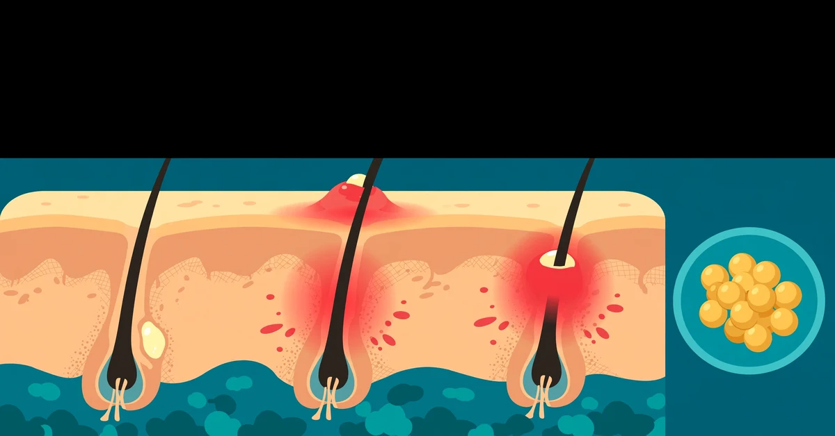
Folliculitis Pictures: What Infected Hair Follicles Look Like on Each Body Area

Hernias by Picture
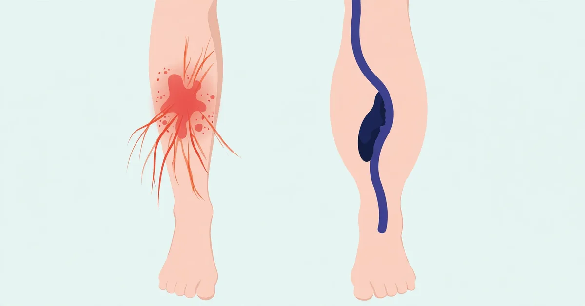
Cellulitis vs DVT: How to Tell a Skin Infection From a Blood Clot in Your Leg

Why Is My Sweat Salty? The Science Behind Sweat
Related Posts

How to Remove and Prevent Moths in Your Home
Aug 16, 2026 • 28 min read
MRSA Pictures: What MRSA Looks Like and When It Becomes Dangerous
Nov 9, 2026 • 5 min read
6 Natural Treatments for Shingles
Sep 8, 2026 • 33 min read
What’s the Best Antiseptic for Open Wounds? Iodine, Peroxides, and More
Aug 15, 2026 • 35 min read
What Causes Peau D’Orange?
Aug 23, 2026 • 31 min read