Pregnancy is an exhilarating period. You’re nurturing a tiny person, eagerly awaiting their arrival, and gearing up for a fresh chapter in your life. It can feel overwhelming at times!
One constant should be a sequence of predictable milestones in your prenatal care.
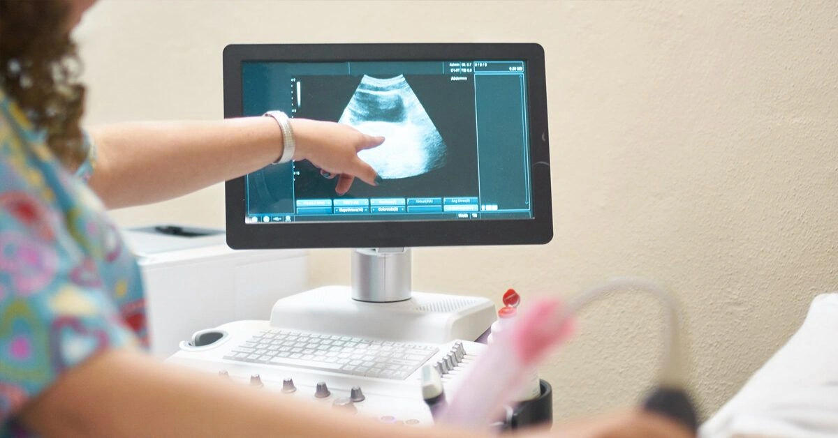
In particular, ultrasounds are pivotal moments when you can catch a glimpse of your developing little one while your physician can confirm that the pregnancy is advancing as it should or identify any critical concerns that might require attention.
Let’s explore what you might anticipate at a 16-week ultrasound.
Reasons you might have a 16-week ultrasound
Although two ultrasounds during a typical pregnancy are common, you might have more — whether due to personal risk factors or your doctor’s preference to monitor the baby’s progress more closely.
A 16-week ultrasound isn’t a standard requirement — but don’t let that worry you! Consider it an opportunity to get additional glimpses of your baby.
Your first ultrasound usually occurs between 8 and 14 weeks to hear a heartbeat and confirm pregnancy while estimating a due date.
The next ultrasound typically happens between 18 and 20 weeks. This scan checks overall fetal development and is sometimes referred to as the “anatomy scan.”
Beyond the two standard ultrasounds, your doctor may arrange extra screenings to:
- verify viability if you missed the earlier heartbeat check
- monitor your pregnancy more closely if you’re at high risk for certain conditions
- check for multiples if there’s a possibility of twins, triplets, or more
- investigate fetal conditions such as heart defects, Down syndrome, or spina bifida
- address any complications you’re experiencing, such as bleeding
How the ultrasound is performed
There’s nothing you need to do to prepare for the 16-week ultrasound.
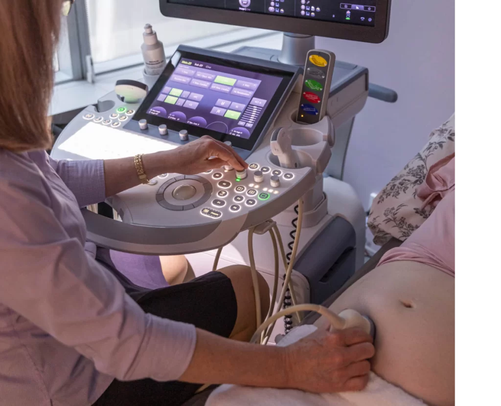
This abdominal scan uses a transducer moved over your abdomen to display 2D images of your developing baby. It’s noninvasive and won’t cause harm to you or your baby, though you might feel some discomfort if the technician needs to press a bit harder at times.
Expect the ultrasound to take about 30 to 60 minutes. During the procedure, your doctor or ultrasound technician — known as a sonographer — will:
- obtain measurements of your baby
- examine the development of their spine
- reconfirm their heartbeat
This is all to ensure that everything is progressing normally.
Depending on the baby’s position, you might be asked to move or turn to allow the technician to view different angles more clearly.
They’ll also check for fetal movement. So, don’t be surprised if your sonographer gently nudges your abdomen to encourage your baby to move if they’re not particularly active during the appointment.
What you might see
During the 16-week scan, you can expect a fully formed but still small baby. If development is on track, you should still be able to see arms, legs, fingers, toes, and even facial expressions during the ultrasound.
At this stage, if you want to know (and if the baby cooperates by assuming the right position), your technician can also attempt to determine the baby’s sex as the external genitalia should be visible in certain positions. However, confirmation may require later verification.
How big your baby is
At 16 weeks, your baby is typically between 4 and 5 inches long and weighs around 5 ounces.
During your 16-week visit, your physician may measure your fundal height, though this is usually started at 20 weeks.
This noninvasive metric assesses the distance in centimeters from the top of your baby bump to the top of your pubic bone. It helps confirm that your baby is growing properly.
Typically, by the time you reach 24 weeks, your fundal height roughly matches your gestational week in centimeters. So, if you’re 27 weeks along, you’d expect a 27-centimeter fundal height reading.
Nevertheless, there’s a margin of error in the measurements. It’s not unusual for the numbers not to align perfectly with your gestational week—especially before 24 weeks—and this also relates to the accuracy of your due date.
Your due date itself is an estimate with its own margin of error. If you had an early ultrasound to estimate your due date, it will usually be more precise.
But the takeaway is this: don’t panic if the baby is measuring a week or so off in either direction. It’s common.
Additional checks your doctor may perform
The 16-week ultrasound, if performed, is a crucial period where your doctor will want to screen for potential developmental abnormalities. They accomplish this by observing movement and taking measurements, as noted above.
While the 16-week appointment is noninvasive, your physician might also recommend drawing blood to run a triple or quadruple screen for potential issues such as neural tube problems, Down syndrome, or additional chromosomal concerns that can be screened via blood tests.
This screening typically occurs between the 15th and 20th week, though screenings conducted between 16 and 18 weeks are said to be most accurate.
If these blood tests indicate a potential problem, your OB may discuss more invasive diagnostic options such as amniocentesis or chorionic villus sampling (CVS). Alternatively, they could suggest further noninvasive prenatal testing.
Although amniocentesis and CVS are highly effective at confirming developmental abnormalities, they carry small risks that can lead to pregnancy complications such as miscarriage. Therefore, physicians prefer to rely on noninvasive measures—like ultrasound—for initial screening.
Determining the sex with ultrasound
If you don’t want surprises, you can often determine your baby’s sex during the 16-week ultrasound. Since your baby’s external anatomy is fully formed, this should be accurate.
However, remember that depending on how your baby is positioned, your physician or the sonographer may not be able to obtain a clear view of their anatomy to confirm the sex.
If the sonographer cannot obtain a clear read, or if you have doubts, you can always ask your doctor to check the baby’s sex as part of a blood screening or schedule a later follow-up ultrasound to confirm.
Expecting twins
Just as with a single pregnancy, if you’re carrying twins, the 16-week ultrasound will show both babies in detail.
However, don’t be surprised if the scan takes longer, as the technician must be meticulous to ensure each baby’s measurements are accurate and properly labeled.
At this stage, each baby should be roughly the same length and size as a singleton.
However, many OBs use a growth chart tailored for twins, since multiples are often smaller at birth than babies from single pregnancies.
Also, it’s not unusual for one twin to be slightly smaller than the other, which is common—only a large discrepancy would raise concern.
The bottom line
The 16-week ultrasound often marks your first serious glimpse of your baby. This milestone helps ease anxiety and makes the pregnancy feel more tangible.
Although noninvasive, this ultrasound is a key step in screening for potential developmental issues and confirming that the baby is growing properly.
Not every pregnancy will include a 16-week ultrasound, but somewhere between 16 and 20 weeks gestation you’ll likely complete this important check.


Leave a Reply
You must be logged in to post a comment.