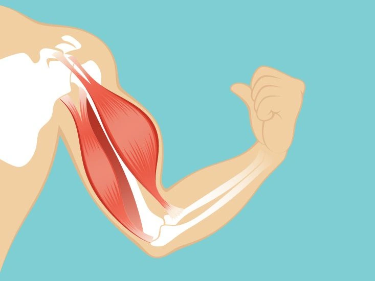
The primary muscle of the chest is the pectoralis major. This broad, fan-like muscle stretches from the armpit to the collarbone and spans across the lower portion of the chest on both sides. The two halves meet at the sternum, commonly called the breastbone.
This muscle facilitates four separate movements at the shoulder joint and also secures the arms to the torso. Tears or strains of this muscle are uncommon, but when they occur they can cause chest pain, bruising, and diminished muscle strength.
Beneath the pectoralis major lies the pectoralis minor. This slender, triangular muscle runs vertically along the upper ribs.
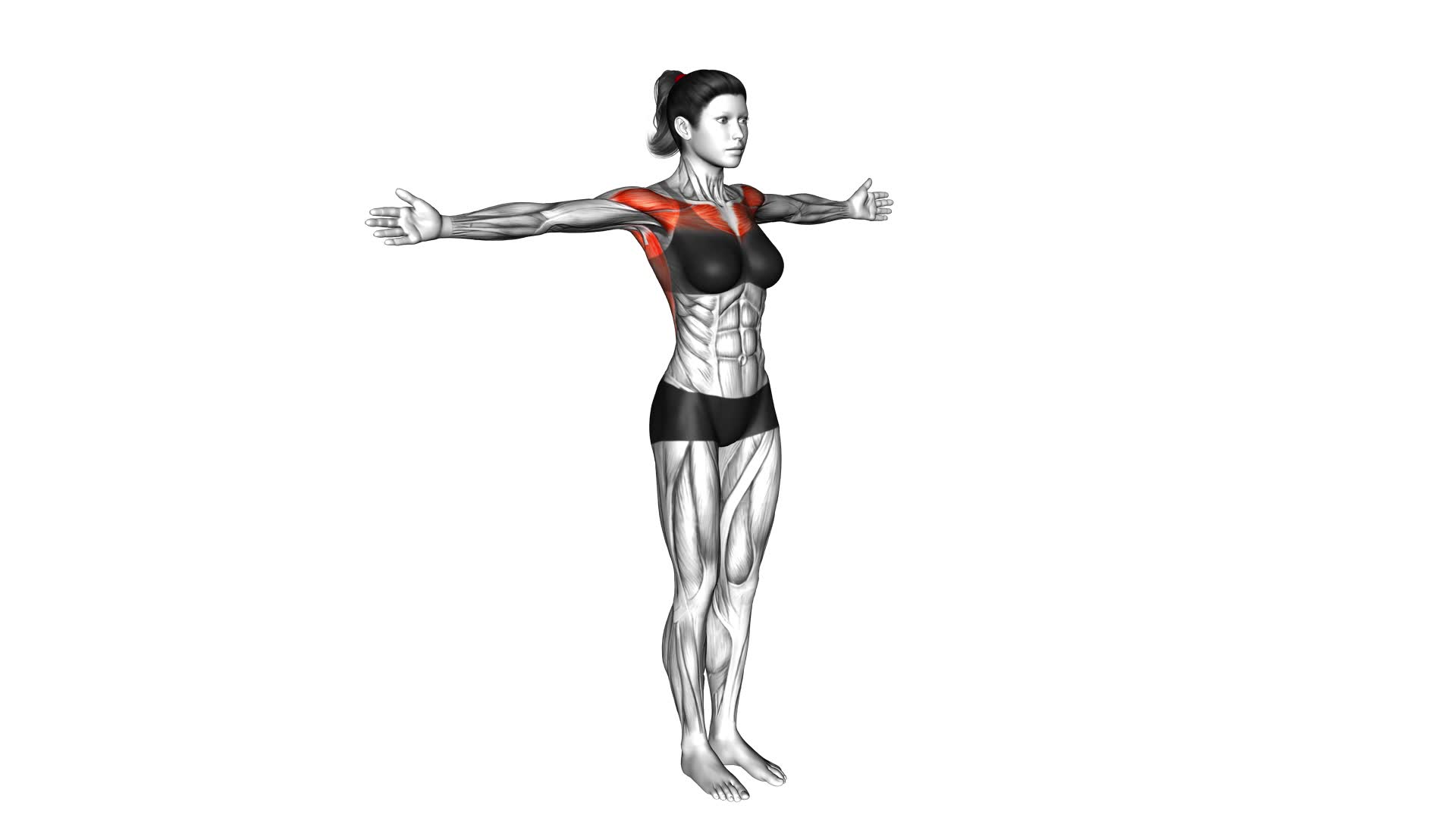
The key muscles of the upper trunk include:
- Trapezius: Spanning the neck, shoulders, and upper back, this muscle enables motion of the shoulders and shoulder blades.
- Rhomboid major: Connected to the scapula, this muscle is one of several that support shoulder motion.
- Infraspinatus: Part of the rotator cuff, it assists in lifting and lowering the arm.
- Teres major: This muscle contributes to rotation of the upper arm.
- Serratus anterior: Positioned along the rib cage, it stabilizes the scapula against the chest wall and helps rotate it upward.
- Deltoid: Giving the shoulder its rounded appearance, the deltoid lifts and rotates the arm.
- Latissimus dorsi: This broad, flat back muscle helps rotate the arms and draw them toward or away from the body.
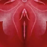
In women, the chest typically contains greater fat deposits than in men, which serve to protect the accessory sex organs — the breasts. These adipose layers help cushion and safeguard the milk-producing glands and their ducts.
The mammary glands produce milk for a newborn after childbirth. Each gland consists of numerous lobules, which are the milk-secreting glands. Lobules connect to ductal lobes, which in turn join with the lactiferous ducts.
The lactiferous ducts form a branching, tree-like system that converges at the nipple. These ducts widen beneath the areola to store milk that accumulates.
Milk exits the body through tiny openings on the nipple. These are variously called milk ducts, mammary ducts, or galactophores.
The mammary glands and surrounding tissues begin to develop as secondary sex structures at the onset of puberty. Nevertheless, the breasts remain functionally inactive until hormonal shifts during pregnancy trigger milk production.
A mother is able to continue producing milk for years provided there is ongoing stimulation through breastfeeding or regular pumping.


Leave a Reply
You must be logged in to post a comment.