
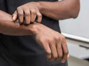
A myxoid cyst is a small, noncancerous lump that appears on a finger or toe, typically close to a nail. It’s also known as a digital mucous cyst or mucous pseudocyst.
The precise origin of myxoid cysts remains uncertain, though they are frequently linked to osteoarthritis. Estimates suggest that 64 percent to 93 percent of people with osteoarthritis develop myxoid cysts.
These cysts most often arise in individuals aged roughly 40 to 70, but they can occur at any age. Women are affected about twice as often as men.
The term myxoid means mucus-like, derived from the Greek for mucus (myxo) and resemblance (eidos). Cyst originates from the Greek word for bladder or pouch (kystis).
What causes myxoid cysts?
Although the exact mechanism isn’t established, there are two main theories.
- One theory is that the cyst arises when synovial tissue around a finger or toe joint deteriorates. This is commonly tied to osteoarthritis and other degenerative joint conditions. Occasionally a tiny bony spur (osteophyte) from degenerating joint cartilage is involved.
- The other theory proposes the cyst develops when fibroblast cells in the connective tissue produce excess mucin (a component of mucus). This form does not involve joint degeneration.
In some instances, notably in people under 30, a finger or toe injury may play a role. A small subset of individuals may develop myxoid cysts from repeated finger motion.
Signs and symptoms
Myxoid cysts are typically:
- small, round or oval lumps
- up to 1 centimeter (cm) in diameter (about 0.39 inch)
- smooth to the touch
- firm or filled with fluid
- not commonly painful, although the adjacent joint may ache from arthritis
- skin-colored or translucent with a red or blue hue and often resembling a “pearl”
- slow to enlarge

These cysts most often form on the dominant hand, typically on the middle or index finger near the nail. Cysts on the toes are uncommon.
If a cyst grows onto the nail it can create a groove or split the nail, and in some instances may lead to nail loss.
Subungual (under the nail) myxoid cysts are unusual and can be painful depending on how they deform the nail.
If you injure a myxoid cyst, it may discharge a sticky fluid. Seek medical attention if the cyst appears infected.
Treatment options
Most myxoid cysts do not cause pain. If the cyst doesn’t bother you cosmetically or functionally, treatment isn’t required and monitoring may be sufficient. Keep in mind that a myxoid cyst rarely disappears spontaneously.
There are numerous treatment options, each with well-documented pros and cons.
Recurrence after treatment is common. Studies of recurrence rates for various therapies exist. Some treatments may:
- leave a scar
- cause pain or swelling
- reduce joint range of motion
If you want the cyst removed, talk to your physician or a specialist about which approach is best. Treatment choices include:
Nonoperative
- Infrared coagulation. Heat is used to cauterize the cyst base. A 2014 literature review reported recurrence rates of 14 percent to 22 percent with this method.
- Cryotherapy. After draining the cyst, liquid nitrogen is applied to freeze and thaw the tissue, aiming to stop further fluid from entering the cyst. Reported recurrence ranges from 14 percent to 44 percent. Cryotherapy can be painful for some.
- Carbon dioxide laser. The laser ablates the cyst base following drainage. This has about a 33 percent recurrence rate.
- Intralesional photodynamic therapy. The cyst is drained, a photosensitizing agent is injected, and laser light is used to destroy the cyst base. A small 2017 study of 10 people reported 100 percent success with no recurrence at 18 months.
- Repeated needling. A sterile needle or blade punctures and drains the cyst; this may be repeated two to five times. Recurrence rates are 28 percent to 50 percent.
- Injection of steroid or sclerosing agents. Various chemicals such as iodine, alcohol, or polidocanol may be injected. This approach shows the highest recurrence: 30 percent to 70 percent.
Surgical
Surgical approaches tend to have high success, with reported rates between 88 percent and 100 percent. For this reason, surgery is often recommended as a primary option.
Surgery removes the cyst and covers the defect with a skin flap that heals over time. The size of the flap depends on the cyst’s dimensions. Surgeons sometimes scrape the joint and remove osteophytes (bony projections from joint cartilage).
At times, dye is injected into the joint to locate and seal the site of fluid leakage. The flap may be sutured, and a splint might be prescribed postoperatively.
Both surgical and nonsurgical strategies rely on producing scar tissue that interrupts the connection between the cyst and the joint, preventing fluid refilling the cyst. Based on treatment of 53 patients, one investigator suggested that this scarring can be achieved without excising the cyst and creating a skin flap.
At-home approaches
You can attempt daily firm compression for a few weeks.
Do not try to puncture or drain the cyst yourself because of the infection risk.
There is anecdotal support for soaking, massaging, and applying topical steroids to help myxoid cysts.
Prognosis
Myxoid cysts are benign. They are not infectious and typically cause no symptoms. They are frequently associated with osteoarthritis in the fingers or toes.
Many treatment options exist, both nonsurgical and surgical, but recurrences are common. Surgical excision generally produces the best outcomes with the lowest recurrence.
If the cyst is painful or cosmetically bothersome, consult your doctor about treatment choices and likely results. Seek prompt medical care if your myxoid cyst shows signs of infection.


Leave a Reply
You must be logged in to post a comment.