Breast Cancer on Ultrasound: How to Read the Pictures and Your Report

- A note on the pictures in this article
- How breast ultrasound makes a picture
- The six features radiologists check on every lump
- 1. Shape
- 2. Margin (the edge of the lump)
- 3. Orientation
- 4. Echo pattern
- 5. Posterior features (what appears behind the lump)
- 6. Calcifications, blood flow and surrounding tissue
- Benign-leaning vs suspicious features at a glance
- What common findings typically look like
- Simple cyst
- Fibroadenoma
- Lipoma
- Invasive ductal carcinoma
- BI-RADS 0 to 6: what your category means
- Ultrasound vs mammography vs MRI: when each is used
- How to read your own ultrasound report, line by line
- What ultrasound cannot do
- When to seek care promptly
- The bottom line
Short answer: On a breast ultrasound, a lump that leans benign usually looks oval, lies flat and parallel to the skin, has smooth and clearly defined edges, and lets sound pass through so the tissue behind it looks brighter. A lump that raises concern usually looks irregular in shape, stands taller than it is wide, has spiky, angled or blurred edges, appears dark (hypoechoic), and casts a shadow behind it. No single feature proves or rules out cancer — radiologists weigh all of these features together, summarise them in a BI-RADS category, and only a biopsy can confirm a diagnosis.
This guide explains, in plain language, what doctors actually look at on breast ultrasound pictures, what each common finding typically looks like, what the BI-RADS numbers 0 to 6 mean for you, how ultrasound compares with mammography and MRI, and how to read your own ultrasound report line by line. It is educational only: it cannot diagnose your lump, and any new breast lump needs an in-person examination and formal diagnostic imaging.
A note on the pictures in this article
Real ultrasound images of breast cancer are medical records, and most are protected by copyright, so websites cannot simply copy the clearest teaching examples from textbooks or clinics. Rather than mislabel a mammogram or an unrelated scan as an ultrasound — a shortcut some pages take — this article uses only ultrasound images with a verified open licence from Wikimedia Commons, and captions each one with exactly what it shows. Where no openly licensed real image exists for a finding, the finding is described in words and in the comparison table below instead of being illustrated with a substitute picture.

How breast ultrasound makes a picture
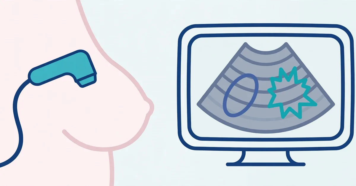
Ultrasound uses high-frequency sound waves, not radiation. A handheld probe sends sound into the breast, and the echoes that bounce back are converted into a live grey-scale picture. Fluid lets sound pass easily and appears black. Solid tissue reflects more sound and appears in shades of grey or white. Fat, glandular tissue, muscle and a lump all return different echo patterns, which is why radiologists describe a mass by its echo pattern: anechoic (no echoes, black), hypoechoic (darker than surrounding tissue), isoechoic (similar brightness) or hyperechoic (brighter than surrounding tissue).
Because the picture is live, the person scanning can press gently, turn the probe, measure the lump in two directions, check blood flow with Doppler, and scan the armpit lymph nodes in the same visit. Ultrasound is especially good at answering one practical question — is this lump fluid-filled or solid? — and at guiding a needle precisely into a lump during a biopsy. RadiologyInfo, the patient site of the Radiological Society of North America and the American College of Radiology, describes breast ultrasound as a standard tool for evaluating a felt lump, clarifying a mammogram finding, and imaging dense breast tissue.
The six features radiologists check on every lump
Breast ultrasound reporting follows a standard vocabulary developed by the American College of Radiology (the BI-RADS lexicon), so that one doctor’s description means the same thing to another. Six features do most of the work.
1. Shape
An oval shape is reassuring and typical of fibroadenomas and cysts. A round shape is neutral; some benign and some malignant masses are round. An irregular shape — neither oval nor round, with an uneven outline — is a suspicious feature, because cancers often grow unevenly into surrounding tissue.
2. Margin (the edge of the lump)
A circumscribed margin means the edge is sharp and you could trace a clean line around the whole lump; this favours a benign cause. Margins that are indistinct (blurred), angular (with sharp corners), microlobulated (with many tiny bumps) or spiculated (with thin lines radiating outward like sun rays) are suspicious. Spiculated margins are among the strongest single warning signs on ultrasound, because they reflect tumour tissue and a fibrous reaction spreading into the tissue around it.
3. Orientation
Benign lumps usually grow along the natural tissue planes, so they lie parallel to the skin and look wider than tall. A mass that is not parallel — taller than wide, growing across the tissue planes toward the skin and chest wall — is a classic suspicious feature. A widely cited Radiology study of solid breast masses found that oval shape, circumscribed margins and a wider-than-tall profile were the features that most reliably characterised masses as benign, while irregular shape and spiculated or microlobulated margins characterised malignant ones.
4. Echo pattern
A simple cyst is anechoic: completely black inside with no internal echoes at all. Most cancers are hypoechoic, darker than the fat around them, and markedly dark masses raise more concern. Hyperechoic (bright) solid masses are uncommon and are usually benign, such as a lipoma made of fat. A mixed, uneven internal pattern is described as heterogeneous and needs closer assessment than a uniform one.
5. Posterior features (what appears behind the lump)
Sound that passes easily through fluid arrives strongly behind a cyst, making the tissue there look brighter — posterior enhancement, a benign sign. Dense, fibrous tumour tissue blocks and scatters sound, leaving a dark column behind the mass — posterior shadowing, a suspicious sign commonly seen with invasive cancers. Some masses show neither, and some show a combination.
6. Calcifications, blood flow and surrounding tissue
Tiny bright specks inside or near a mass can represent microcalcifications, which are more often a mammogram finding but, when seen on ultrasound inside a suspicious mass, add concern. Doppler imaging may show increased or disorganised blood flow in cancers, although many benign lumps also have blood flow, so this is a supporting clue rather than a deciding one. Pulling-in or distortion of the tissue around a mass, skin thickening, and abnormal armpit lymph nodes all raise the level of concern.
Benign-leaning vs suspicious features at a glance
| Feature | More often benign | More suspicious |
|---|---|---|
| Shape | Oval | Irregular |
| Margin | Circumscribed, smooth | Spiculated, angular, microlobulated, indistinct |
| Orientation | Parallel, wider than tall | Not parallel, taller than wide |
| Echo pattern | Anechoic (simple cyst); uniform | Markedly hypoechoic; heterogeneous |
| Behind the mass | Posterior enhancement | Posterior shadowing |
| Calcifications | Coarse, in a known benign lump | Microcalcifications in or near a solid mass |
Features are weighed as a set, not in isolation. A lump with one mildly unusual feature but an otherwise classic benign pattern may simply be watched, while a lump stacking several suspicious features will be biopsied promptly. There are also honest exceptions in both directions: a few cancers look round and tidy, and a few benign conditions — such as fat necrosis after an injury, radial scar or sclerosing adenosis — can look irregular and alarming. That overlap is exactly why imaging sorts lumps into risk categories instead of giving a final answer, and why tissue sampling exists.

What common findings typically look like
Simple cyst
The textbook benign picture: round or oval, completely black inside, thin smooth wall, brighter tissue behind it (posterior enhancement) and no blood flow inside. A lump meeting all of these strict criteria is classified as benign and usually needs no treatment unless it is painful or very large, in which case it can be drained with a fine needle. If a fluid collection contains debris, thick walls or a solid component, it is no longer called “simple” and is managed more cautiously.
Fibroadenoma
The most common benign solid lump, especially in younger women. On ultrasound it is typically an oval, wider-than-tall solid mass with smooth or gently lobulated, well-defined edges, a uniform grey interior, and sometimes a thin bright rim (a pseudocapsule of compressed tissue). Older fibroadenomas can develop coarse “popcorn” calcifications. A classic-looking fibroadenoma is often monitored; a new, growing or atypical one is biopsied to be certain, because a minority of cancers can mimic this tidy appearance.
Lipoma
A benign lump of fat. On ultrasound it is usually oval, parallel and soft-looking, with brightness similar to or brighter than the surrounding fat and fine internal lines. Lipomas are typically left alone unless they grow or cause symptoms.
Invasive ductal carcinoma
The most common breast cancer, and the one behind most “suspicious” ultrasound descriptions. Its typical picture is the mirror image of the benign pattern: an irregularly shaped, dark (hypoechoic) solid mass, taller than wide, with spiculated, angular or microlobulated margins, posterior shadowing, sometimes microcalcifications, increased Doppler blood flow, and distortion of the tissue around it. Not every invasive cancer shows every feature, and small or special-type cancers (for example mucinous cancers) can look deceptively smooth — another reason biopsy, not appearance, makes the diagnosis.
BI-RADS 0 to 6: what your category means
At the end of the report, the radiologist assigns a BI-RADS (Breast Imaging Reporting and Data System) assessment category. It is not a cancer stage and, except for category 6, it is not a diagnosis — it is a standard statement of how concerned the radiologist is and what should happen next. The same scale is used for mammography and breast MRI.
| Category | Meaning | Usual next step |
|---|---|---|
| 0 | Incomplete — more imaging or old images for comparison are needed before a final assessment | Additional views, ultrasound or comparison with prior studies |
| 1 | Negative — nothing abnormal seen | Routine screening as advised |
| 2 | Benign finding, such as a simple cyst | Routine screening; no treatment for the finding itself |
| 3 | Probably benign — chance of cancer is 2% or lower | Short-interval follow-up imaging, commonly at 6, 12 and 24 months, to confirm stability |
| 4 | Suspicious — split into 4A (low, over 2% up to 10%), 4B (moderate, over 10% up to 50%) and 4C (high, over 50% up to under 95%) | Biopsy is usually recommended; most category 4 results still turn out benign |
| 5 | Highly suggestive of malignancy — at least 95% likelihood | Biopsy and prompt specialist referral; treatment planning follows the tissue result |
| 6 | Known, biopsy-proven cancer | Imaging used to plan or monitor treatment |
Two categories cause the most anxiety and deserve plain words. BI-RADS 3 does not mean “possible cancer” in the worrying sense: it means the appearance is almost certainly benign, and time — proving the lump does not change — is being used as the test instead of a needle. Keep those follow-up appointments; the safety of the approach depends on completing them. BI-RADS 4 does not mean cancer is likely in every case: the category spans a wide range, most biopsied category 4 lumps are benign, and the biopsy is how the question gets settled.
Ultrasound vs mammography vs MRI: when each is used
Ultrasound is usually the first imaging test for a felt lump in younger women and in pregnancy, the go-to test for telling a cyst from a solid lump, a useful add-on in dense breasts, and the standard way of guiding core-needle biopsies. It uses no radiation and is widely available, but it is operator-dependent and can miss microcalcifications that mammography catches.
Mammography (including 3D tomosynthesis) remains the standard screening test for most women of screening age and the best test for microcalcifications and subtle architectural distortion. When a lump is found in a woman over about 30, a diagnostic mammogram and an ultrasound are often done together, because each sees things the other can miss. If you are working out where lumps tend to occur and what a new lump needs, our guide to where breast cancer lumps are usually found covers locations and next steps.
Breast MRI is the most sensitive of the three and is reserved for specific situations: screening women at very high inherited risk, clarifying conflicting findings, assessing the extent of a known cancer, or checking implants. It produces more false alarms than the other tests and is not a routine first step for a new lump. The U.S. Centers for Disease Control and Prevention and the National Cancer Institute both emphasise that screening and diagnostic choices depend on age, risk and the finding itself — which is why a clinician, not a picture on the internet, should direct your work-up.
How to read your own ultrasound report, line by line
- Indication / history: why the scan was done (for example, “palpable lump, right breast”). Check it matches your symptom and side.
- Technique: which breast and areas were scanned, sometimes including the armpit. Targeted scans focus on the lump; whole-breast scans survey everything.
- Location: given by side, clock position and distance from the nipple (for example, “10 o’clock, 4 cm from nipple”). This is the address of the finding, used to re-find it at follow-up or biopsy.
- Size: measured in up to three dimensions, in millimetres or centimetres. Size matters for follow-up comparison, but a small lump is not automatically safe and a large one is not automatically cancer.
- Description words: shape, margin, orientation, echo pattern and posterior features — the six features explained above. A cluster of benign words (oval, circumscribed, parallel) reads very differently from a cluster of suspicious words (irregular, spiculated, taller-than-wide, shadowing).
- Impression / assessment: the summary and the BI-RADS category. This is the single most important line for what happens next.
- Recommendation: routine screening, follow-up ultrasound in 6 months, or core-needle biopsy. If the recommendation and the category seem to conflict, ask — you are entitled to an explanation in plain language.
For broader context on what breast cancer pictures of different types can and cannot show, see our overview of pictures of breast cancer lumps, and for one specific type, our explainer on HER2-positive breast cancer.
What ultrasound cannot do
Ultrasound sorts lumps by risk; it does not name them. It cannot reliably detect every microcalcification, its quality depends on the equipment and the operator, and some cancers imitate benign lumps while some benign lumps imitate cancer. A reassuring scan also does not overrule a lump that you and your clinician can still feel and that is growing — persistent, unexplained palpable lumps deserve escalation even after a normal scan. Only examination of tissue under a microscope (pathology from a biopsy) confirms whether a lump is cancer and, if so, which type.
When to seek care promptly
Book a prompt appointment for any new lump or thickening, a lump that is growing, skin dimpling or redness, new nipple inversion or discharge (especially bloody), a lump in the armpit, or a breast change that does not settle after one menstrual cycle. Do not wait for pain — most early breast cancers are painless. The NHS advises seeing a GP promptly about any new breast lump or change; earlier assessment gives the widest range of options whatever the cause turns out to be.
The bottom line
On ultrasound, benign-leaning lumps are oval, parallel, smooth-edged and let sound through; suspicious lumps are irregular, taller than wide, spiky or blurred at the edges, dark, and shadow-casting. Radiologists combine those features into a BI-RADS category that states the next step, and biopsy — not any picture — makes the diagnosis. Use this guide to understand your report and to ask sharper questions, not to judge your own scan.
Medical disclaimer: This article is for general education only and is not medical advice. It cannot diagnose any lump from a description or an image. Any new, persistent or changing breast lump needs an in-person clinical examination and formal diagnostic imaging arranged by a qualified healthcare professional.
Frequently Asked Questions
No. Ultrasound sorts lumps into risk levels based on shape, margin, orientation, echo pattern and what appears behind the mass, summarised in a BI-RADS category. Only a biopsy, with tissue examined under a microscope, can confirm or rule out cancer.
It most often appears as a dark (hypoechoic), irregularly shaped solid mass that is taller than wide, with spiculated, angular or blurred margins and shadowing behind it. Not every cancer shows all of these features, and some look deceptively smooth, which is why suspicious or uncertain lumps are biopsied.
A simple cyst is round or oval, completely black inside (anechoic), thin-walled and smooth, with brighter tissue behind it (posterior enhancement) and no internal blood flow. A lump meeting all of these criteria is classified as benign (BI-RADS 2).
BI-RADS 3 means “probably benign”: the estimated chance of cancer is 2% or lower. The usual plan is follow-up imaging at about 6, 12 and 24 months to prove the finding is stable, instead of an immediate biopsy. Keeping those follow-up appointments is essential.
A biopsy is recommended. Category 4 covers a wide suspicion range (4A low, 4B moderate, 4C high) and most category 4 biopsies turn out benign. Category 5 means at least a 95% likelihood of malignancy on imaging, but the diagnosis still comes from the biopsy result, not the scan.
They answer different questions. Ultrasound is better for telling a fluid-filled cyst from a solid mass, is usually first for a felt lump in younger women, and guides biopsies. Mammography is the standard screening test and the best at showing microcalcifications. For a new lump after about age 30, the two are often done together.
Read Next

5 Ways to Reduce Stress-Related Tension in Your Neck and Shoulders

What Is Saturated Fat? Sources, Daily Limits and Smarter Swaps

7 Ways to Remove Dead Skin from Your Feet
Blood Sugar Level Chart: What Your Fasting, After-Meal and Random Readings Mean

6 Resistance Band Exercises for Shoulders
Dacryocystitis
Related Posts
Melanoma Pictures: A Visual Guide to What Melanoma Looks Like
Nov 10, 2026 • 35 min read
Ultrasounds Aren't Typically Used to Detect Cervical Cancer: Learn Why
Aug 31, 2026 • 33 min read
Is the Pain in My Tailbone Cancer?
Aug 21, 2026 • 21 min read
Is It Safe to Mix Naproxen and Acetaminophen?
Aug 18, 2026 • 20 min read
How to Find a Hearing Aid Provider That Accepts Medicaid: A Step-by-Step Guide
Nov 9, 2026 • 14 min read