Tonsillitis Pictures: A Visual Guide to Swollen and Infected Tonsils

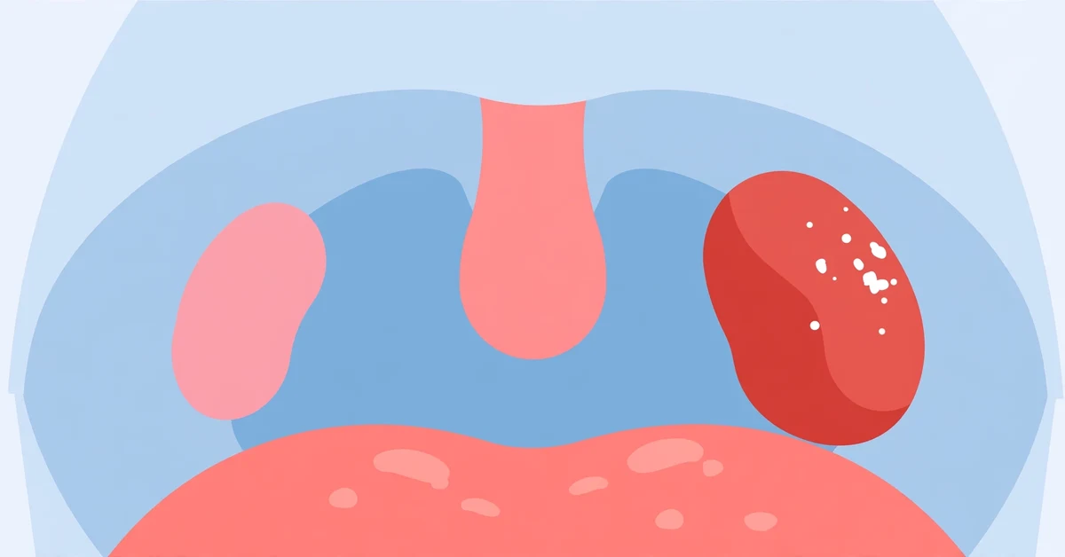
- What healthy tonsils look like
- What tonsillitis looks like at a glance
- Viral tonsillitis: red and swollen, usually without pus
- Bacterial (strep) tonsillitis: white exudate and palate spots
- How to read a tonsil photo: what each feature means
- Viral vs. bacterial tonsillitis at a glance
- Peritonsillar abscess: the one-sided warning picture
- Not tonsillitis: tonsil stones and other look-alikes
- How to check your own tonsils — and take a photo a doctor can use
- Why pictures cannot diagnose tonsillitis
- When to see a doctor — and when to go urgently
- How tonsillitis is treated
- The bottom line
Tonsillitis announces itself visually more than most infections. Open your mouth in front of a mirror during a bad sore throat and you may see two swollen, angry-red tonsils, sometimes coated with white or yellow patches. That is exactly why so many people search for tonsillitis pictures: they want to know whether what they are looking at matches the real thing, and whether it looks viral or bacterial. According to MedlinePlus , tonsillitis is an inflammation of the tonsils — the two oval pads of lymph tissue that sit on either side at the back of the throat — most often caused by a virus, and sometimes by bacteria such as group A streptococcus.
This guide walks through real clinical photographs of healthy and infected tonsils and explains, feature by feature, what you are seeing. One warning before the photos: no picture can diagnose tonsillitis or prove strep throat. A photo can tell you how urgently to get checked, but only an examination and, when needed, a throat swab can confirm the cause. The CDC is explicit that doctors cannot diagnose strep throat by looking at the throat alone — a rapid strep test or throat culture is required.
What healthy tonsils look like
To judge an abnormal photo, you first need the normal baseline. Healthy tonsils are pink — roughly the same color as the rest of the mouth — and sit inside shallow pockets on either side of the throat, behind the folds of tissue called the tonsillar pillars. They are roughly symmetrical, their surface is smooth or lightly grooved, and there is no coating, no white spotting, and no swelling pushing them toward the middle. Size alone is a poor alarm bell: tonsils are naturally large in children, look biggest between about ages 4 and 7, and shrink after puberty. Big but pink, calm, and symmetrical is usually just anatomy.

Use the labels in this photo to orient yourself in the pictures that follow. The dangling structure in the center is the uvula, hanging from the soft palate. The tonsils flank it left and right, and the tongue sits below. When you check your own throat, these are the landmarks to find first — and the uvula matters later in this article, because its position is one of the most important warning signs a photo can show.
What tonsillitis looks like at a glance
Compared with the healthy baseline, tonsillitis changes three things: color, size, and surface. The tonsils turn a deeper, brighter red. They swell — mildly at first, and in severe cases enough to meet in the middle, a finding clinicians call “kissing tonsils.” Doctors grade tonsil size on a scale from 1+ (just visible) to 4+ (touching in the midline). And the surface changes: it may look glossy and streaked with mucus, dotted with white or yellow patches, or sprinkled with tiny red spots on the palate above.

The look is only half the story. Tonsillitis usually arrives with a sore throat that makes swallowing hurt, fever, bad breath, and tender, swollen glands in the neck. Ear pain is common too — the throat and ear share nerve pathways, so an inflamed tonsil often makes a perfectly healthy ear ache (our guide to ear ache with a sore throat explains how that referred pain works). In young children who cannot describe symptoms, drooling, refusing food, and unusual fussiness may be the visible clues.
Viral tonsillitis: red and swollen, usually without pus
Most tonsillitis is viral — caused by the same families of viruses behind common colds, flu, and COVID-19. In photos, viral tonsillitis typically shows tonsils that are red and moderately swollen, often with visibly engorged blood vessels on their surface, but without the thick white or yellow coating of a bacterial infection. The whole back of the throat tends to look generally inflamed rather than focused on the tonsils alone.
The company it keeps is the giveaway. Viral tonsillitis usually travels with cough, a runny or blocked nose, hoarseness, sneezing, or red, watery eyes — symptoms that point away from strep. Some viruses leave distinctive visual signatures: coxsackievirus (herpangina and hand, foot and mouth disease) raises small blisters or ulcers on the tonsils, soft palate, and back of the throat. The NHS notes that viral tonsillitis typically settles on its own within about three to four days with rest, fluids, and pain relief.
There is one viral exception every picture guide should flag: infectious mononucleosis (caused by the Epstein–Barr virus) can coat the tonsils in a heavy white-grey layer that looks exactly like bacterial tonsillitis. If a “strep-looking” throat comes with crushing fatigue, swollen glands all around the neck, and symptoms dragging past a week, mono belongs on the list — and only a blood test can separate it from strep, because the pictures genuinely overlap.
Bacterial (strep) tonsillitis: white exudate and palate spots
When tonsillitis is bacterial, the usual culprit is group A streptococcus — the same germ behind strep throat. Its signature visual feature is exudate: patches, streaks, or a near-complete coating of white to pale-yellow pus sitting in the tonsils’ grooves and folds. The tonsils beneath are typically a vivid, almost raw red and substantially swollen.

The second strep clue is easy to miss because it is not on the tonsils at all. Look at the soft palate — the smooth roof of the mouth just above them. In strep throat it often shows petechiae: pinpoint red spots, like tiny broken blood vessels scattered across the palate. Combined with fiery redness of the whole back of the throat, this is one of the most recognizable strep patterns in a photograph.

The symptom pattern reinforces the picture. Strep tends to start suddenly, with a severe sore throat and fever, and — importantly — usually without a cough. Tender glands at the front of the neck are typical. In children, headache, stomach pain, and nausea are common, and some develop the fine, sandpapery rash of scarlet fever. The Cleveland Clinic emphasizes that because viral and bacterial tonsillitis can look so similar, testing — not appearance — decides who gets antibiotics.
How to read a tonsil photo: what each feature means
When you compare your throat with the photos above, work through these features one at a time. Each one shifts the odds; none of them, on its own, settles the diagnosis.
| What you see | What it usually means |
|---|---|
| White or yellow patches in the tonsil grooves (exudate) | Points toward a bacterial cause — or mononucleosis. Uncommon in a simple viral cold. |
| Deep, bright, almost raw redness | Active inflammation. With palate spots and no cough, it strengthens the case for strep. |
| Tiny red spots on the soft palate (petechiae) | A classic strep clue, produced by inflamed, fragile capillaries. |
| Both tonsils swollen about equally | The typical pattern of ordinary viral or bacterial tonsillitis. |
| One side much more swollen, uvula pushed to the other side | Suspect a peritonsillar abscess until proven otherwise — this needs urgent care. |
| Small blisters or shallow ulcers at the back of the throat | Usually viral — herpangina or hand, foot and mouth disease (coxsackievirus). |
| White chunks sitting in tonsil pockets, tonsil itself calm | Probably tonsil stones, not tonsillitis (see the section below). |
| A thick grey membrane coating the tonsils | Uncommon. Mono can do this; any tough grey membrane warrants same-day medical assessment. |
Viral vs. bacterial tonsillitis at a glance
| Feature | Viral tonsillitis | Bacterial (strep) tonsillitis |
|---|---|---|
| Typical look | Red, swollen tonsils; generally inflamed throat | Vivid red, swollen tonsils, often much larger |
| White coating | Usually none (mono is the exception) | White/yellow exudate is common |
| Soft palate | May be red; ulcers in some viruses | Pinpoint red spots (petechiae) are a classic sign |
| Cough and cold symptoms | Common — cough, runny nose, hoarseness | Usually absent |
| Fever | None or low-grade | Often above 101°F (38.3°C) |
| Onset | Gradual, with cold symptoms | Sudden, severe sore throat |
| Treatment | Supportive care only | Antibiotics — after a positive strep test |
Treat this table as a weighting tool, not a verdict. The features overlap — which is precisely why guidelines from the CDC and Mayo Clinic route the decision through a swab test rather than a photograph.
Peritonsillar abscess: the one-sided warning picture
One complication changes both the picture and the urgency. A peritonsillar abscess (quinsy) is a pocket of pus that collects in the tissue beside one tonsil, usually as tonsillitis worsens rather than improves. Its photograph looks different from everything above: the swelling is overwhelmingly on one side, the soft palate on that side bulges downward and forward, and the uvula is pushed across to the opposite side, like a curtain shoved sideways.

The picture usually comes with a matching set of symptoms: severely one-sided throat pain, a muffled voice (classically described as sounding like speaking with a hot potato in the mouth), difficulty opening the mouth fully, drooling because swallowing is too painful, and bad breath. A peritonsillar abscess is not a wait-and-see problem. It needs same-day urgent or emergency care, where treatment is drainage of the abscess plus antibiotics; left alone, the swelling can threaten the airway.
Not tonsillitis: tonsil stones and other look-alikes
The most common false alarm in tonsil photos is not an infection at all. Tonsil stones (tonsilloliths) are small, white-to-yellow chunks of trapped debris and bacteria that harden inside the tonsil’s natural pockets. In a photo they look like white spots — but the tonsil around them is its normal pink color, neither red nor swollen, and the main complaint is bad breath rather than a painful, feverish sore throat. If your white spots sit in the tonsil crevices without redness, swelling, or fever, our dedicated guide to what tonsil stones look like will help you confirm the difference.
Two other look-alikes are worth knowing. Oral thrush paints white patches across the tongue, cheeks, and throat that can be wiped off, leaving sore red skin beneath — it is a fungal infection, not tonsillitis, and favors babies, denture wearers, and people using steroid inhalers. And hand, foot and mouth disease covers the back of the throat with small ulcers while adding a rash on the hands and feet. The pattern around the spot — not just the spot — is what separates these conditions.
How to check your own tonsils — and take a photo a doctor can use
A good look beats a bad photograph, and both start with light. Wash your hands, stand in front of a mirror, and shine a phone flashlight or a second phone’s torch into your mouth. Open wide, stick your tongue out, and say “ahh” — this lifts the soft palate and exposes the tonsils. If the tongue blocks the view, press it down gently with the handle of a clean spoon, on the front two-thirds only; pushing further back triggers the gag reflex and ends the exam.
For a photo, tap the screen to focus on the tonsils, keep the light source close to the camera, and take two or three shots. Then read the picture the way the decoder above does: compare the two sides for symmetry, look for coating in the grooves, check the palate for pinpoint spots, and note where the uvula sits. Be aware of the limits: bathroom lighting can make a normal throat look redder than it is, you cannot see behind the tonsils or down the throat, and children rarely cooperate long enough for a clear shot. A reassuring photo does not rule infection out, and an angry-looking one does not prove strep is in.
Why pictures cannot diagnose tonsillitis
Clinicians do not diagnose from appearance alone either. They weigh visual findings alongside features known as the Centor criteria — fever, tonsillar exudate, tender front-of-neck glands, and absence of cough — and then, when strep is plausible, they confirm it with a rapid strep antigen test, backed up by a throat culture in children and teenagers when the rapid test is negative. The reason for the discipline is that the overlap is real: mono imitates strep, strep sometimes produces no exudate at all, and most sore throats are viral and gain nothing from antibiotics. A photograph — yours, or one scored by an app — can prioritize how fast you seek care. It cannot replace the swab.
When to see a doctor — and when to go urgently
Some findings in a throat picture, combined with how you feel, mean urgent, same-day, or emergency care:
- Trouble breathing, noisy breathing, or a feeling that the throat is closing
- Drooling, or being unable to swallow your own saliva
- One-sided swelling with the uvula pushed off-center, or a new muffled voice
- Difficulty opening the mouth fully
- Severe one-sided throat pain, or pain spreading into the neck
- Signs of dehydration — very little urine, dizziness, dry mouth
- A baby or young child with fever who is not drinking
Book a prompt appointment instead if a sore throat is not improving after three to four days, fever runs above 101°F (38.3°C), you can see white patches on the tonsils, or episodes keep coming back. The NHS advises medical review when tonsillitis symptoms persist beyond about four days or keep recurring — repeated, documented episodes are also what starts a conversation about surgery.
How tonsillitis is treated
Treatment follows the cause, which is why the test matters. Viral tonsillitis is managed with supportive care: rest, plenty of fluids, warm saltwater gargles, throat lozenges, and acetaminophen or ibuprofen for pain and fever. Most cases improve within three to four days and clear within a week. Confirmed strep tonsillitis is treated with antibiotics — penicillin or amoxicillin are first-line — taken for the full prescribed course; antibiotics shorten the illness modestly, make you non-contagious after about 24 hours, and, most importantly, prevent complications such as rheumatic fever. A peritonsillar abscess needs drainage plus antibiotics, not antibiotics alone.
When tonsillitis returns again and again — the commonly used threshold is around seven episodes in one year, five per year for two years, or three per year for three years — an ENT specialist may discuss removing the tonsils. If you are weighing that decision for yourself or your child, our guide to the tonsil removal age limit covers who qualifies and what recovery involves.
The bottom line
In pictures, tonsillitis is a pattern: tonsils that are redder, bigger, and rougher-surfaced than the healthy pink baseline — sometimes coated white, sometimes not. White exudate plus fever plus no cough tilts toward strep and deserves a swab test, not a guess. A one-sided bulge with the uvula pushed off-center is the one picture that should never wait: that is an abscess until a clinician proves otherwise. Use photos to decide how fast to get care — and let examination and testing make the diagnosis.
This article is general information only and is not medical advice. If you are worried about your breathing, swallowing, or a rapidly worsening sore throat, seek urgent care rather than comparing photos.
Frequently Asked Questions
Tonsillitis shows tonsils that are redder and more swollen than normal, sometimes meeting in the middle of the throat. The surface may be glossy and streaked, coated with white or yellow patches (exudate), or accompanied by tiny red spots on the soft palate. Healthy tonsils, by contrast, are pink, symmetrical, and uncoated. The overall pattern — color, size, surface, and symmetry — matters more than any single feature.
No. White or yellow exudate is a classic strep sign, but mononucleosis (mono) can coat the tonsils just as heavily, and some white spots are not infection at all — tonsil stones sit in the tonsil pockets without making the tonsil red or swollen. Because the causes look alike, a rapid strep test or throat culture, not the photo, decides whether antibiotics are needed.
Only as a probability. Bacterial (strep) tonsillitis more often shows thick white exudate, pinpoint red spots on the palate, high fever, and no cough; viral tonsillitis is usually red and swollen without pus and arrives with cough, runny nose, or hoarseness. The overlap is large enough — especially with mono — that doctors confirm strep with a swab rather than trusting appearance.
A peritonsillar abscess makes the swelling strikingly one-sided: the tissue beside one tonsil bulges down and inward, and the uvula is pushed across to the opposite side. It usually comes with severe one-sided pain, a muffled voice, trouble opening the mouth, and drooling. This is an urgent problem — it needs same-day care for drainage and antibiotics, because the swelling can endanger the airway.
If the white or yellow chunks sit inside the tonsil’s pockets while the tonsil itself stays pink, normal-sized, and painless, they are most likely tonsil stones — hardened debris whose main symptom is bad breath. Tonsillitis makes the whole tonsil red, swollen, and painful, usually with fever. When there is no redness, swelling, or fever, stones are the better explanation.
Viral tonsillitis typically improves within three to four days and clears within about a week with rest, fluids, and pain relief. Confirmed strep tonsillitis starts improving within 24 to 48 hours of antibiotics, though the full course must be finished. A sore throat that keeps worsening after four days, or one-sided swelling at any point, should be reassessed in person.
Seek urgent care for trouble breathing, drooling or inability to swallow saliva, difficulty opening the mouth, one-sided swelling with the uvula off-center, a muffled voice, or signs of dehydration. Book a prompt appointment if a sore throat is not improving after three to four days, fever exceeds 101°F (38.3°C), you can see white patches, or episodes keep recurring.
Read Next

Guide to Baby Wearing: Benefits, Safety Tips, and How To
Cellulitis vs DVT: How to Tell a Skin Infection From a Blood Clot in Your Leg

Hiya Vitamins Review By a Mom Who Is a Dietitian

Midlife Crisis or Midlife Myth? What to Know About Going ‘Over the Hill’

How to Use a Sled Push to Build Power, Speed, and Endurance

What Causes an Enlarged Uterus and How Is It Treated?
Related Posts

How to Use a Warm Eye Compress to Treat Styes and Other Conditions
Sep 9, 2026 • 3 min read
Natural Pain Relievers
Sep 4, 2026 • 8 min read
Hello Wisp Review: Birth Control, UTIs, STIs, and More
Sep 29, 2026 • 36 min read
Pregnancy Rib Pain: Causes, Prevention, Remedies
Sep 19, 2026 • 19 min read
How to Check for Yeast Infections: The 4 Best At-Home Vaginal pH Tests
Sep 24, 2026 • 21 min read