Not Just Sticks and Toes
Okay. Quick pop quiz: If your foot was a gadget, what would it be? If you said “old potato”… well, I get it—most of us totally overlook these hardworking things. But try this instead: Imagine your foot as a tiny, high-tech suspension bridge—full of springs, shock absorbers, hinges, and electrical wires. I used to call mine “paddle boards” until I learned what’s really going on in there.
Trust me, once you actually get a peek at the parts of the foot diagram, everyday stuff starts making so much more sense—like why you stub a toe and swear you’ll never walk again, or why a new pair of sneakers can have you strutting (or limping) for days.
Map Out the Mystery
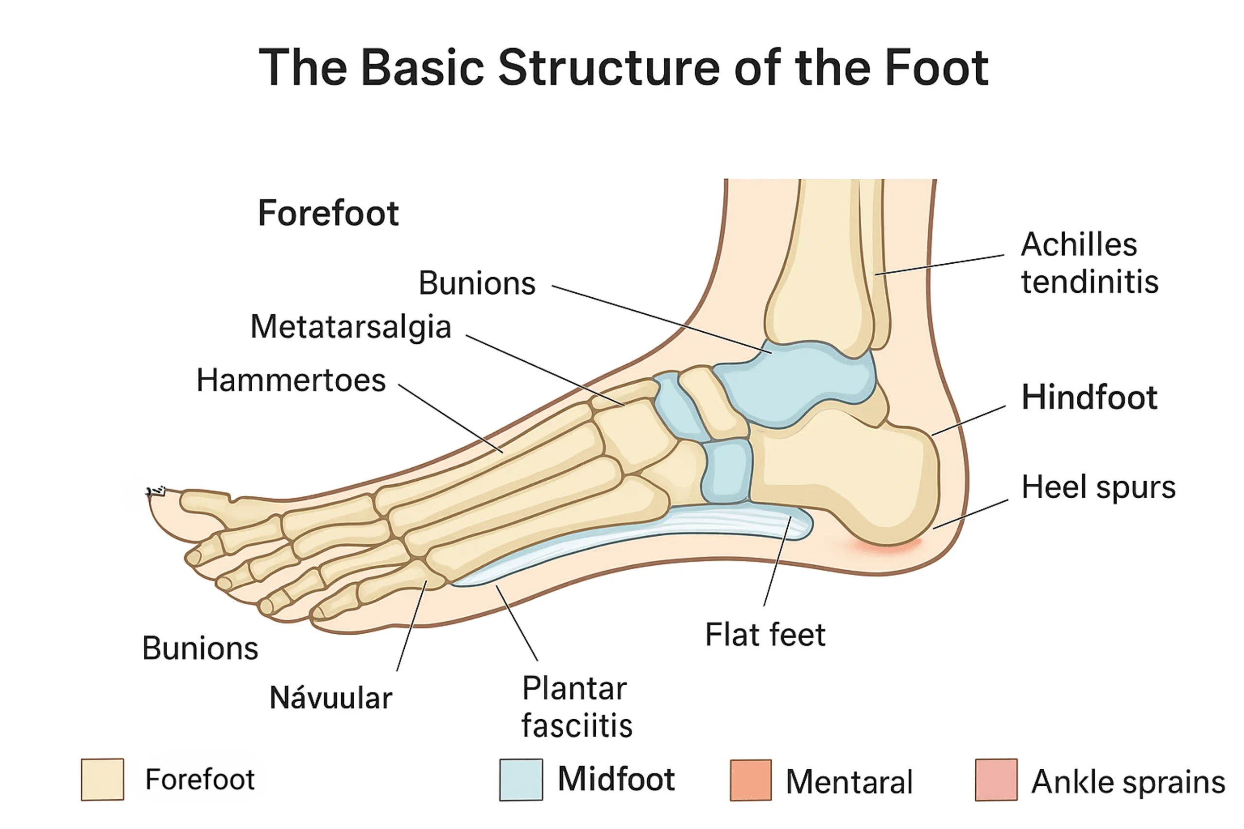
So, what’s really inside your foot? Let’s break it out together. (Don’t worry, we’ll keep it breezy and jargon-free—if I can get it, you definitely can!) The foot is clever: divided into three main zones—hindfoot, midfoot, and forefoot. Think of these like the “neighborhoods” of your one-foot town.
Starting in the Back: Hindfoot Hangout
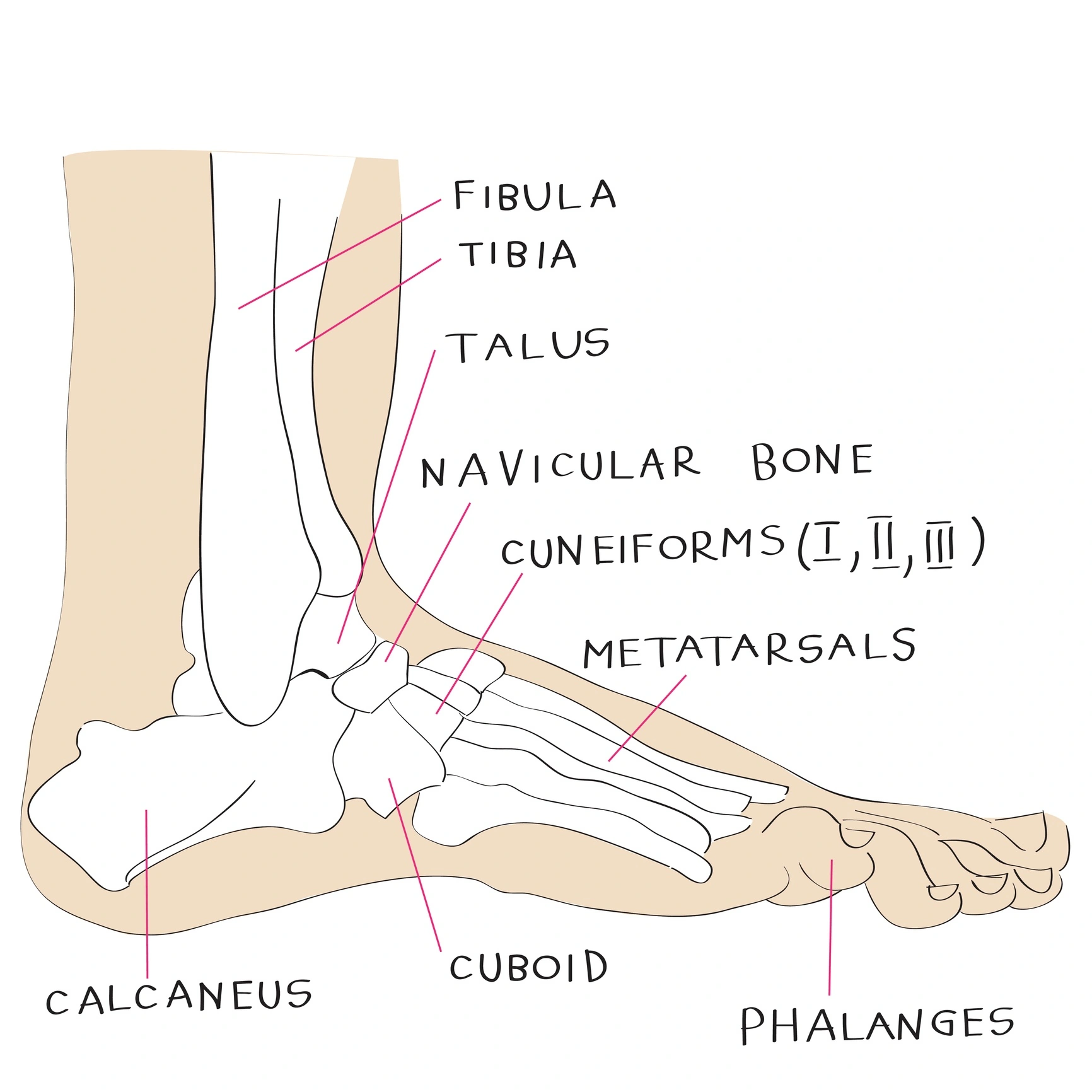
This is your anchor, your sturdy base camp. The so-called “hindfoot” is made up of the talus (the main ankle bone) and the rockstar calcaneus (your heel). If you’ve ever missed a curb or landed hard from a jump—yep, the calcaneus took that hit for you. Talus is like the “silent ninja” of your gait, letting bones in your ankle move freely while still handling a chuck of your weight. (Mine cracked once. It’s… not fun. Wear good shoes, friends.)
Quick Table: Hindfoot in Action
| Bone | Real-Life Comparison | Job in Your Foot |
|---|---|---|
| Calcaneus | Your foot’s bumpers (like car shock absorbers) | Absorbs impact, supports weight |
| Talus | Bicycle ball joint | Connects foot to leg, helps with flex & balance |
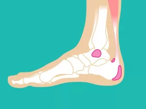
Curious how these look from below? Check out a labeled Foot anatomy bottom diagram—the heel and midfoot components are way more visible from this angle than you might think.
Midfoot: The Flexible Bridge
Let’s shuffle a little forward. Right in the middle sits the midfoot—basically, the arch of your foot and center of shock absorption. In fancy terms, it holds the navicular, cuboid, and three charmingly named cuneiforms (medial, intermediate, lateral). Picture these bones locking together like Lego bricks—except, you know, holding up your whole body weight as you sprint for the bus.
If you’ve ever had arch pain or “that one ache just ahead of your heel,” you know the midfoot. I have this friend—a dancer—who didn’t even know about cuneiform bones until she strained one… now she stretches her arches with the devotion of a yoga teacher. (Lesson: don’t skip foot warm-ups.)
Why Does My Arch Always Ache?
Midfoot bones do a few things—form the springy bridge, help your foot flex and twist, and spread the force every time you step. Think of your foot like a fancy bridge—they’re the suspension cables! And those cuneiforms? They line up with your toes so you don’t topple over whenever you miss a stair.
- Navicular: Kinda boat-shaped, anchors the inside of your arch
- Cuboid: Sits outside, stabilizes and keeps your foot from wobbling
- Cuneiforms: Three in a row, let you roll through your step
Table: Midfoot MVPs
| Bone | Nickname | Main Job |
|---|---|---|
| Navicular | Boat bone | Boosts arch strength |
| Cuboid | Outer stabilizer | Stabilizes outer foot, disperses forces |
| Cuneiforms | The trio | Link arch to toes, help roll the foot |
For a labeled blueprint, find a Foot parts name diagram—so helpful for connecting those wacky medical names to the bones you actually feel when you flex.
Front and Center: Forefoot Moves
We’ve reached the “business end”—the area that hits the floor every time you take a step. The forefoot packs real power. Here you’ll find the five metatarsals (think “toe stalks”—one for each toe), your 14 phalanges (all your toe bones), and a pair of tiny sesamoids hiding under your big toe joint. Yes… humans have pea-sized “knee caps” on their toes. Who knew?
Have you ever stubbed your pinkie toe and seen stars? That’s a metatarsal or phalange hollering at you. These bones are smaller, but don’t underestimate them—they take on huge loads. According to research on foot anatomy, some runners put tons of force on a single forefoot bone with every stride.
Toe Bones: Tiny Yet Mighty
This is the launcher of your steps. The first metatarsal (under your big toe) is the strongest—it does most of the work during that final push-off. The smaller toes help with balance, jostling, and “grip” when you walk on weird surfaces.
And those sesamoids? They’re hidden like surprise bonus features, easing pressure and helping your big toe bend. (Fun fact: Dancers sometimes fracture these—my ballet friend’s X-ray looked like she had lentils under her toe!)
Quick Table: Forefoot Breakdown
| Bone | Count per foot | Fun Fact |
|---|---|---|
| Metatarsals | 5 | Shape your forefoot, key to push-off |
| Phalanges | 14 | Big toe has only 2 (proximal & distal), others have 3 |
| Sesamoids | 2 | Embedded in tendons; act like toe “knee caps” |
Curious what your foot looks like underneath? The Foot anatomy bottom diagram makes every ridge and bump clear. Plus, you can show off the next time someone complains about their cramps. (Might not win you friends at parties, but it’s good trivia!)
Beyond the Bones: Connectors and Controllers
Bones are cool, but they’d be nothing without the connectors—tendons, ligaments, nerves. If the bones are the frame, these are the wires, pulleys, and “zip ties” that keep you standing, walking, leaping, skipping… even dancing (or limping to your fridge at midnight).
So, How Does It All Move?
Over 100 tendons and a sea of ligaments and nerves do the hard work. Take the Achilles tendon—it stretches from the back of your calf down to your heel. That’s why “Achilles injury” hurts like heck: it flexes every time you move, run, hop, or chase your dog down the street.
- Plantar fascia: Keeps your arch springy. When irritated? Hello, dreaded plantar fasciitis (I’ve been there—ice, roll, repeat).
- Peroneal tendons: Outside of your ankle—keep you steady and stop your foot from rolling.
- Tibialis anterior: Lifts your toes so you don’t trip. Stairs, anyone?
- Medial plantar nerve: Feeds your big toe & inner sole with “messages” so you can sense every pebble or Lego brick—ouch.
For a full map of these, poke through a handy Foot parts name guide. Sounds nerdy—feels empowering. (You’ll finally know what your podiatrist is muttering about.)
Your Feet in Real Life: Why Knowing the Parts Matters
Let’s be honest. Most of us don’t give our feet a second thought… until they hurt, or stop working, or—let’s be real—smell funky! But knowing the parts of the foot diagram isn’t just “anatomy for anatomy’s sake.” It helps you make better decisions every day. Picking shoes, deciding when to rest, figuring out which part to ice? It’s all easier when you know what’s what.
Ever had weird pain after a long walk? Now you can ask: “Is this my midfoot? Metatarsal? Did I overdo it and anger my plantar fascia—again?” Seriously. Once you know which “neighborhood” is grumpy, you can look up quick remedies or find better shoes that support your particular arch or avoid squishing your toes.
Here’s a story: My cousin thought all foot pain was just “old age.” Turns out, his pain was coming from a cranky navicular bone—midfoot, near the arch. One appointment and a good arch support later, he’s walking his dog like a champ.
If you’re the type who loves visuals, taking a few minutes to study the Foot parts name chart—especially with a good side-by-side from the Foot anatomy bottom—is super helpful. You’ll finally recognize what those little dots and arrows at the doctor’s office are actually pointing at. (Bonus: You’ll sound really smart the next time foot pain comes up in conversation… which, let’s face it, is more often than you’d think.)
Making Peace with Your Parts
You made it. You’ve peeked inside your heel, traced your arch, and counted (probably with toes wiggling) all those little bones at the end of your foot. That’s… a lot. But now you know—the parts of the foot diagram aren’t just textbook trivia. They’re a map to less pain, better performance, and more confident walks on every wacky sidewalk you’ll ever cross.
So, what now? Next time your feet feel off, try to pinpoint the “neighborhood.” Pamper your arches, pick your sneakers with smarts, and maybe—just maybe—give your toes a little stretch and love before you hit the ground running. If you’re ever curious or confused, head back to that Foot parts name or wander through the underside with the Foot anatomy bottom guide. Your feet will thank you—and so will every step you take.
Now… when’s the last time you did something nice for your feet? Maybe today’s the day. Even a minute of attention goes a long way toward happier, comfier walks—and hey, maybe fewer “ow ow OW!” moments, too. I’m cheering for you (and your ten trusty toes) every step of the way.


Leave a Reply
You must be logged in to post a comment.