The foot represents the lowest portion of the human leg and plays a crucial role in everyday movement. Understanding Foot anatomy bottom helps explain how humans are able to walk, run, jump, and climb with stability and coordination. The unique design of the foot, combined with the body’s built-in balance mechanisms, allows for both strength and flexibility during motion.
This intricate structure is composed of more than 100 tendons, ligaments, and muscles that coordinate the movement of nearly three dozen joints. While the overall arrangement of bones in the foot resembles that of the hand, the foot is engineered to support body weight. As a result, it is sturdier and less mobile, prioritizing stability over dexterity.
Bones That Form the Foundation
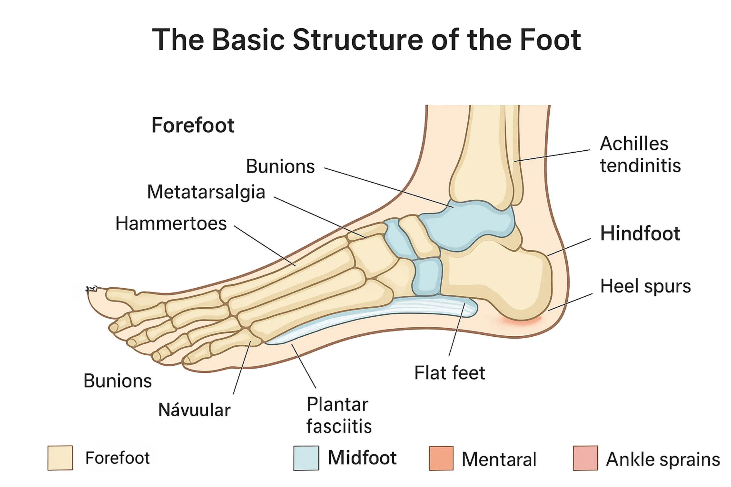
The largest bone in the foot is the calcaneus, which forms the heel. This bone angles upward to connect with the tarsal bones, while the remaining foot bones extend forward and downward to create the full framework of the sole. In discussions of Foot anatomy bottom, the calcaneus is often highlighted because it absorbs much of the impact generated during walking and running.
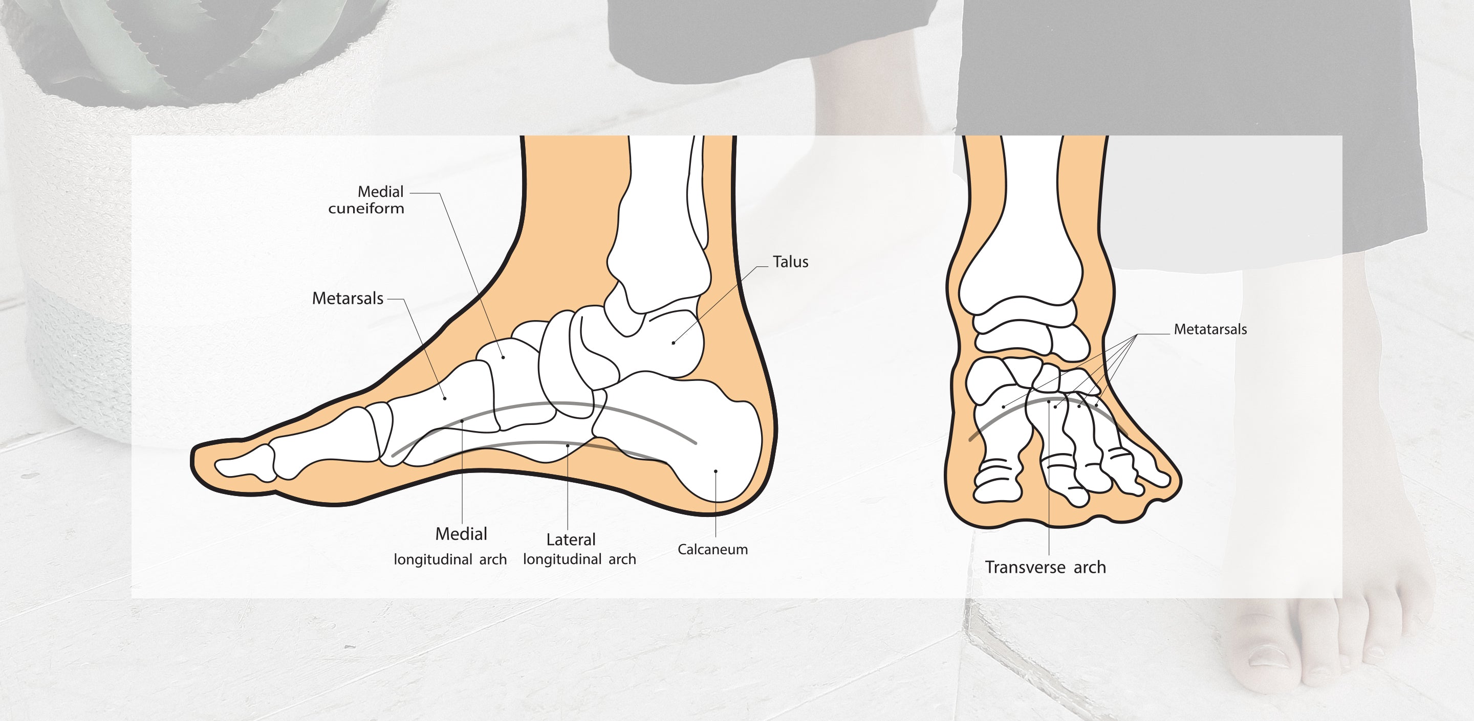
Beneath the connection point of these bones lie the arches of the foot. These three curved structures along the bottom of the foot help distribute weight efficiently, making movement smoother and reducing strain on the body. The arches include the medial arch, lateral arch, and fundamental longitudinal arch. They are formed by the specific angles of the bones and reinforced by tendons, which connect muscles to bones, and ligaments, which connect bones to one another.
If you want a clearer visual understanding of how these bones align, reviewing a detailed Parts of the foot diagram can provide helpful anatomical context.
Bone Groups and Their Organization
The bones in the foot are arranged into three primary groups: the tarsal bones, metatarsal bones, and phalanges. Together, these structures form the toes and the broader front portion of the foot. Knowing each Foot parts name can improve your understanding of how injuries or medical conditions affect specific regions.
Additional bones contribute to ankle structure and connectivity, including:
- Tibia
- Fibula
- Talus
- Cuneiforms
- Cuboid
- Navicular
These bones work together to create a stable yet adaptable base that supports body weight while permitting controlled motion. The talus, in particular, plays a central role in transferring weight from the leg to the foot.
Muscles, Tendons, and Ligaments
Although many muscles responsible for larger foot movements originate in the lower leg, the foot itself contains a complex network of smaller muscles. These intrinsic muscles enable precise movements, assist with balance, and help the foot adapt to uneven surfaces. The coordinated action of tendons and ligaments ensures that the arches maintain their shape while allowing necessary flexibility during walking.
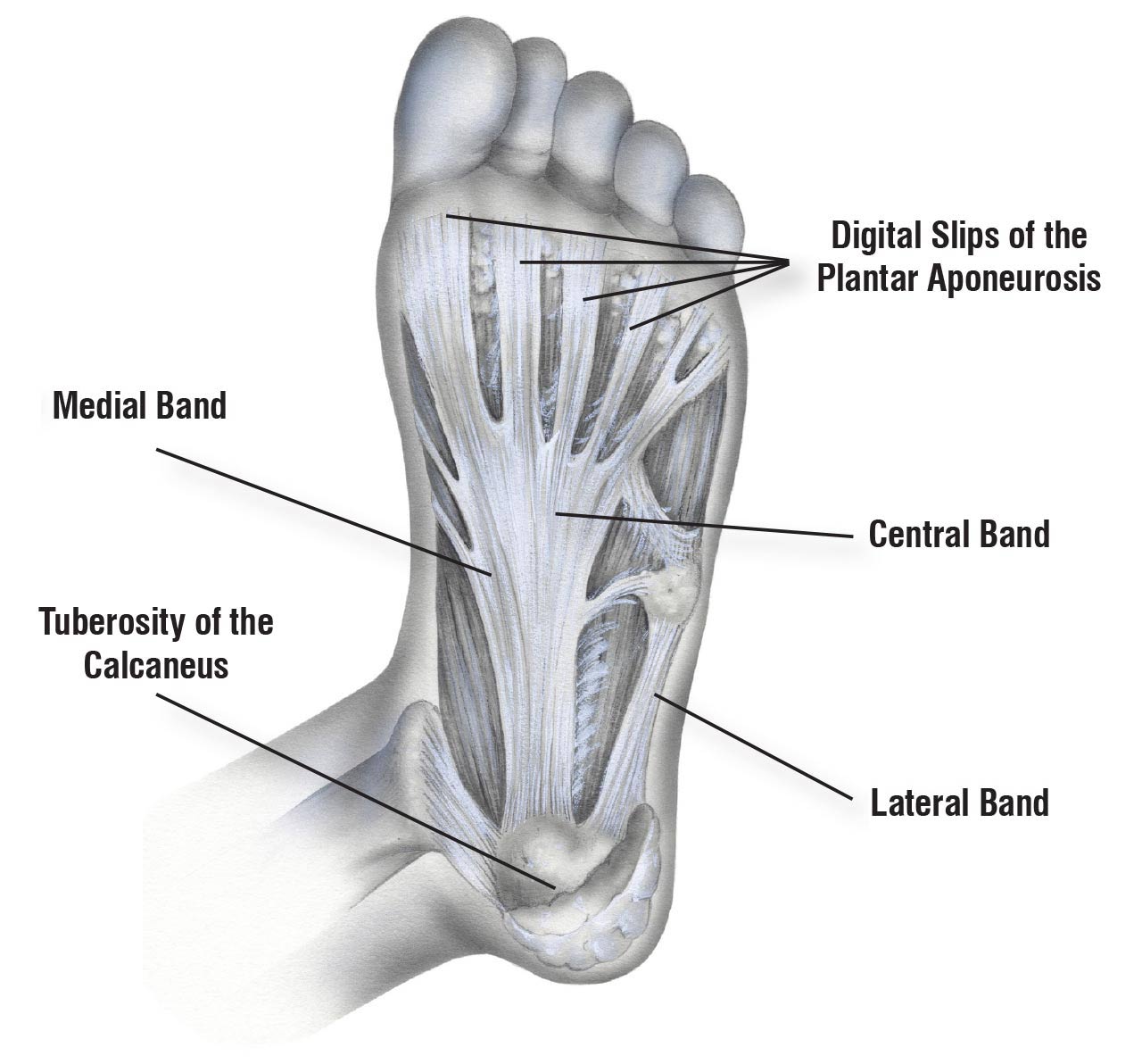
The bottom of the foot contains soft tissues that act as natural shock absorbers. These tissues cushion impact and protect bones and joints. When functioning properly, this integrated system minimizes stress on the knees, hips, and spine.
The positioning and mechanics of the feet significantly influence overall posture and spinal alignment. Problems affecting Foot anatomy bottom can extend beyond localized discomfort, sometimes contributing to back pain or joint strain. Improper footwear is a common contributing factor. Shoes that fail to support the natural arches or disrupt weight distribution may increase the risk of discomfort and injury.
Wearing supportive footwear that complements the foot’s natural alignment can help maintain healthy arches and reduce unnecessary pressure. Individuals who spend long hours standing or walking may benefit from cushioned soles and proper arch support to protect the structures on the bottom of the foot.
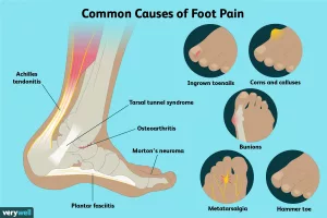
Common Foot Problems
Because the feet bear the entire weight of the body, they are susceptible to a variety of conditions. Some issues arise from overuse, while others may result from infections, systemic diseases, or improper biomechanics.
- Foot pain
- Athlete’s foot
- Plantar warts
- Gout (a type of arthritis)
- Plantar fasciitis (heel pain)
- Stress fractures
- Diabetic foot ulcers
Understanding and Preventing Foot Conditions
Foot pain can stem from structural strain, inflammation, or injury to the bones and soft tissues. Plantar fasciitis (heel pain) occurs when the plantar fascia becomes irritated, often due to repetitive stress or inadequate arch support. Stress fractures may develop from overtraining or sudden increases in physical activity.
Infections such as athlete’s foot and plantar warts affect the skin on the bottom of the foot, particularly in warm, moist environments. Gout (a type of arthritis) can cause intense joint inflammation, frequently affecting the big toe. Meanwhile, diabetic foot ulcers are serious complications that require prompt medical attention, especially in individuals with diabetes.
Preventive measures include wearing well-fitted shoes, maintaining a healthy body weight, stretching the calf muscles and plantar fascia, and practicing good foot hygiene. Regular inspection of the feet is especially important for individuals with diabetes, as early detection of skin changes or sores can prevent complications.
A strong understanding of Foot anatomy bottom empowers individuals to recognize symptoms early and take steps to protect foot health. Since the feet serve as the foundation of the body, caring for them supports overall mobility, balance, and long-term well-being.


Leave a Reply
You must be logged in to post a comment.