
Although the embryo remains quite tiny at this stage, an 8-week ultrasound can often confirm a heartbeat.
Early prenatal visits can feel surreal — particularly if this is your first time expecting. These initial checkups are typically focused on establishing a health baseline for pregnancy and ensuring development is on track.
One important early milestone is the 8-week ultrasound. So why is an ultrasound scheduled so soon, and what should you anticipate during this appointment? Below we cover those questions and more.

What occurs during the 8-week ultrasound?
Even though a pregnancy test can return positive roughly two weeks after conception, it may take longer for the tiny cluster of cells to show physical changes that confirm normal progression. In particular, clinicians want to verify that the embryo has a heartbeat — a definitive sign of viability.
In some instances, a heartbeat can be detected as early as 6 weeks. After a positive at-home test, call your provider to find out if an ultrasound is warranted.
Transvaginal versus abdominal scans
When most people picture an ultrasound, they imagine a technician sweeping a probe across a gel-coated belly — an abdominal ultrasound. Early scans usually take less than 30 minutes.
A transvaginal ultrasound, by contrast, involves inserting a probe into the vagina to obtain a closer view of the embryo and surrounding structures; it’s frequently used in early pregnancy.
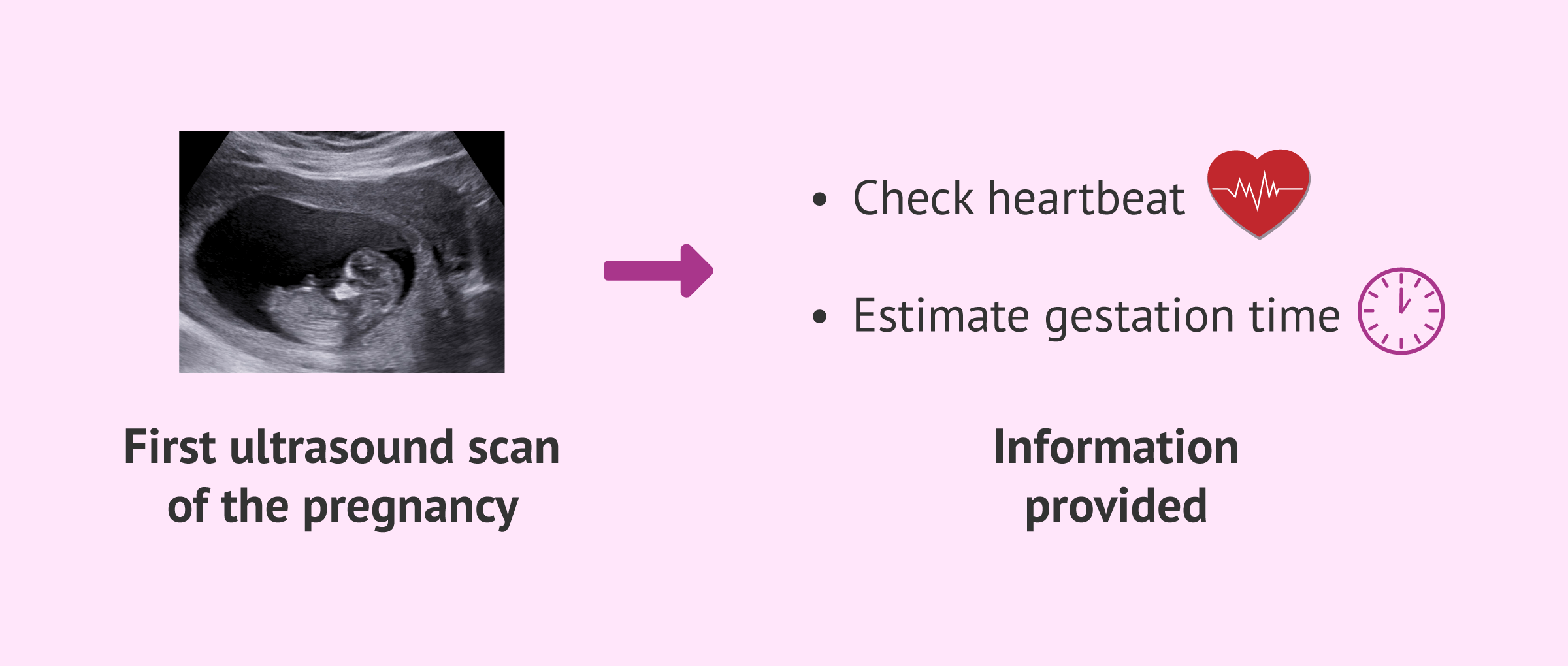
Besides checking for a heartbeat, the clinician will look for features like the gestational sac and the fetal crown-rump length. These measurements help establish gestational age and refine your due date.
What will you likely see at 8 weeks?
This appointment offers your first real glimpse at your developing baby. Don’t expect fine details yet — the image will be simple.
You’ll probably notice a small shape resembling an oblong bean; if you’re carrying twins, two such shapes may be visible. The head remains proportionally large compared with the body.
The gestational sac — the fluid-filled cavity surrounding the embryo — will be visible, and within it the yolk sac, a round structure that supports the embryo early on. Depending on the scan and equipment, you may also hear the heartbeat.

What is the clinician checking for?
The primary goals of the 8-week scan are to confirm intrauterine pregnancy, estimate a due date, and verify the fetal heartbeat. Your provider will look for markers such as a gestational sac and a fetal pole to ensure the pregnancy is located inside the uterus; seeing more than one sac could be your first clue of multiples.
Once pregnancy is confirmed, the next step is to update your expected due date. An earlier estimate might have been provided based on the first day of your last menstrual period, but that method can be inaccurate because cycles vary. The usual dating calculation uses the LMP, subtracts three months, and adds one year and seven days, but individual cycles can shift this estimate.
An ultrasound allows measurement of the embryo to determine gestational age and a more precise due date. The clinically recommended approach in early pregnancy is the crown-rump length (CRL) measurement, which is generally accurate to within about 5–7 days during the first trimester (see guidance).
If the embryo or heartbeat aren’t seen
Sometimes neither the embryo nor a heartbeat is visible — and that doesn’t always indicate a poor outcome. One common reason is that conception occurred later than assumed; if ovulation happened later, the scan might simply be too early to detect expected structures.
Other factors, like large uterine fibroids or anatomical variations, can make imaging more challenging.
However, in some cases the absence of an intrauterine embryo could point to an ectopic pregnancy, where implantation has occurred outside the uterus. Other possibilities include a blighted ovum, when the gestational sac forms but the embryo does not develop, or an early miscarriage.
Your clinician can interpret the findings for your specific situation and advise when it may be appropriate to attempt pregnancy again if that is desired.
What’s happening with the embryo at eight weeks?
The first trimester is a time of rapid development. During week eight, many foundational systems and structures are forming.
At about 8 weeks, the embryo is roughly the size of a kidney bean and may measure nearly half an inch. While it won’t yet resemble the newborn you’ll eventually meet, its features are becoming more recognizably human.
Arm and leg buds are present and beginning to take shape into fingers and toes, though they may appear webbed now. Bones, muscles, and skin are developing, though the skin remains translucent at this stage. The embryo is active and continually moving even though you won’t feel it yet.
How you might feel during week eight
The first trimester can bring a flood of symptoms in addition to emotional highs. Around 8 weeks, common physical symptoms often intensify. Typical complaints include:
- fatigue
- tender or sore breasts
- morning sickness
- nausea that can persist through the day
- difficulty sleeping
- frequent urination
- heartburn
The takeaway
Once you receive a positive pregnancy test, contact a healthcare professional to determine when you should be evaluated and potentially scanned. An early ultrasound is commonly scheduled to confirm pregnancy, refine your due date, and ensure the embryo has a healthy heartbeat.
Your 8-week visit may include either a transvaginal or an abdominal ultrasound; both are low-risk and can provide the first real look at your baby. Keep in mind that because this is still very early, it’s possible that a heartbeat or the embryo itself won’t be visible yet.
For more information on what to expect at early scans and timing, especially around the second trimester, you may find this 16 week ultrasound resource helpful.


Leave a Reply
You must be logged in to post a comment.