Key takeaways
- Melanoma may show up in a range of colors — not only the classic brown or black — and it can form anywhere on the body, although it’s seen more often on the trunk in men and on the legs in women.
- There are multiple types of melanoma, each with unique features; superficial spreading melanoma is the most frequently diagnosed, while nodular melanoma tends to be more aggressive.
- Routine skin self-exams are essential for catching changes early. Any mole that becomes asymmetrical, develops uneven borders, changes in color, increases in diameter, or evolves over time should be assessed promptly by a dermatologist.
Melanoma is among the less common varieties of skin cancer, yet it is the most dangerous due to its ability to spread (metastasize) to distant organs if not identified and treated early.
In 2022, approximately 99,780 individuals are expected to receive a melanoma diagnosis, and more than 7,600 deaths are projected. Melanoma rates continue to increase, making awareness and early detection especially important.
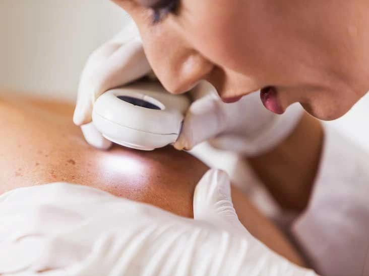
Reviewing Skin cancer pictures early stages can help you recognize suspicious changes and understand what melanoma may look like before it advances. Below, you’ll learn how to identify early warning signs and when to seek medical care.
Pictures of melanoma
Melanoma is a form of cancer that develops in melanocytes, the pigment-producing cells of the skin. It may also be referred to as malignant melanoma or cutaneous melanoma.
While many melanomas appear brown or black, they can also present as pink, tan, red, blue, or even white lesions. Because of this variation, reviewing Skin cancer pictures early stages across different skin tones can be particularly helpful.
There are four primary types of melanoma, and their appearance can differ depending on factors such as skin tone and location. Many melanomas are flat or only slightly raised and often display multiple colors with uneven or jagged borders.
The tumor’s thickness is measured using the Breslow measurement, also known as Breslow‘s depth. This measurement plays a key role in determining the stage of the melanoma and guiding treatment decisions.
Melanoma can arise on any part of the skin, but it is more likely to begin on the trunk (chest and back) in men and on the legs in women. The face and neck are also frequent sites of involvement.
Most moles are benign and remain stable over time. However, individuals with numerous moles are more likely to develop melanoma. Warning signs include noticeable changes in a mole’s size, contour, color, or border irregularity.
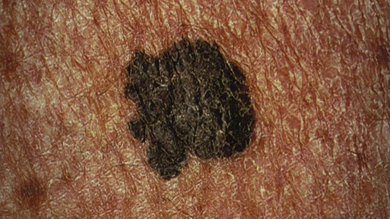
Superficial spreading melanoma
Superficial spreading melanoma is a type of skin cancer that initially grows horizontally within the upper layers of the skin before penetrating deeper layers.
It represents about 70 percent of all melanoma diagnoses, making it the most common subtype. Features and symptoms to watch for include:
- a flat or slightly elevated lesion, often with an irregular shape and uneven borders, sometimes arising from an existing mole or appearing as a new one
- colors such as brown, black, tan, red, blue, or white — often darker than the person’s usual skin tone
- gradual changes that may develop over months or even years
For comparison with other non-melanoma skin cancers, you can also explore Basal cell carcinoma Stages pictures to understand how early lesions differ in appearance.
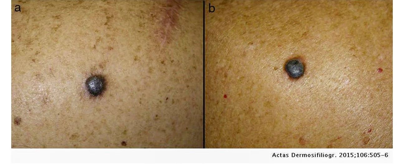
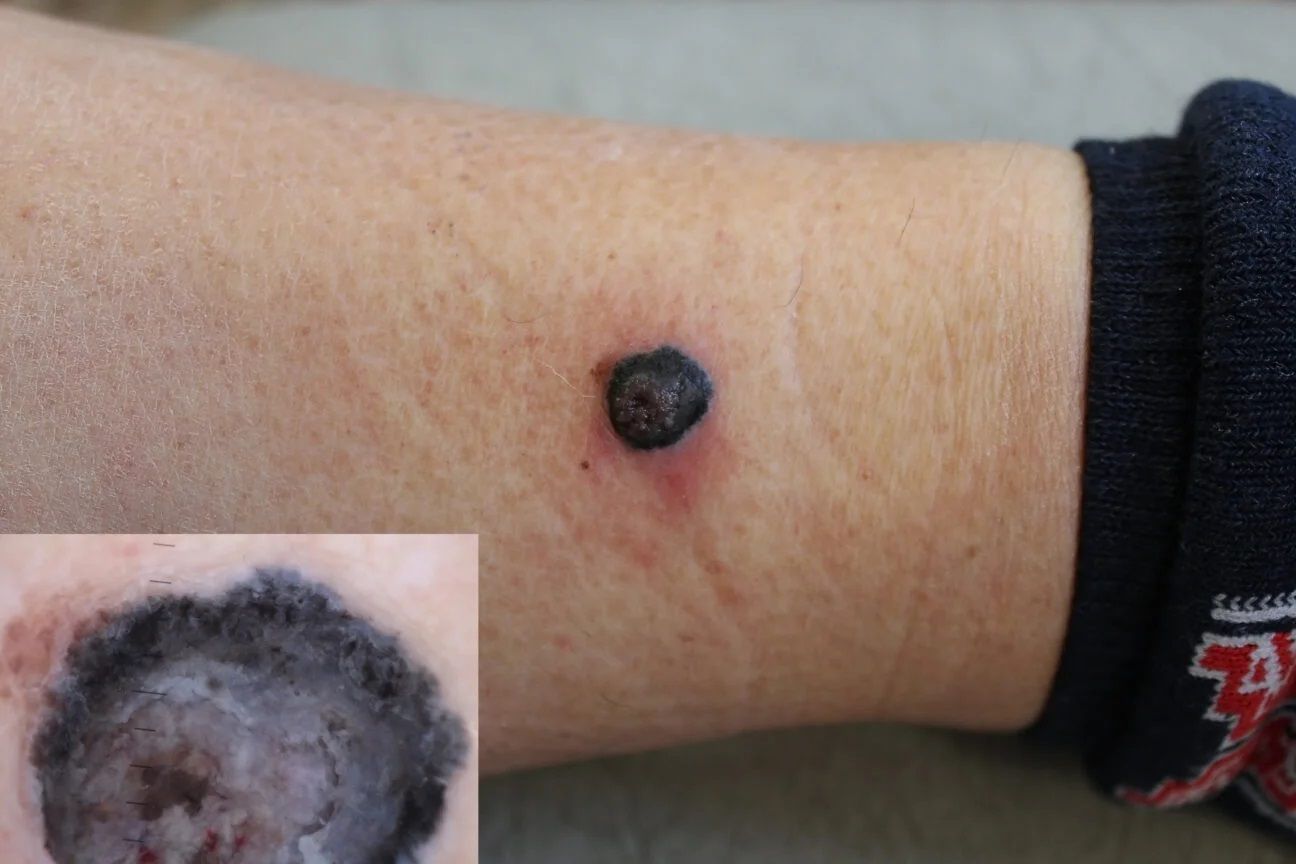
Nodular melanoma
Nodular melanoma is considered one of the most aggressive forms of skin cancer because it tends to grow vertically into deeper layers of the skin early in its course. Signs and characteristics include:
- a firm, raised bump
- a blackish-blue, dark brown, or reddish-blue color (sometimes similar to surrounding skin)
- rapid growth in size and shape, particularly over 2 to 3 weeks
Advanced cases may resemble those seen in Pictures of advanced basal cell carcinoma, although the underlying cancer type differs.
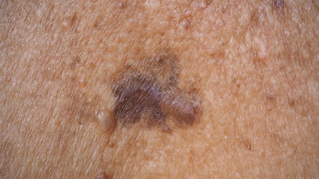
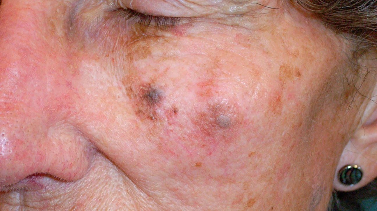
Hutchinson’s melanotic freckle (aka lentigo maligna melanoma)
Hutchinson’s melanotic freckle is an invasive skin cancer that arises from lentigo maligna, which is a melanoma in situ confined to the uppermost skin layer. Although lentigo maligna is not initially invasive, it can become cancerous and progress to lentigo maligna melanoma. Concerning features include:
- a large, flat or slightly elevated brown or black patch resembling an age spot or freckle
- a smooth surface with an irregular outline
- a brown coloration that may occasionally appear red, pink, or white depending on skin tone
- a diameter typically at least 6 millimeters
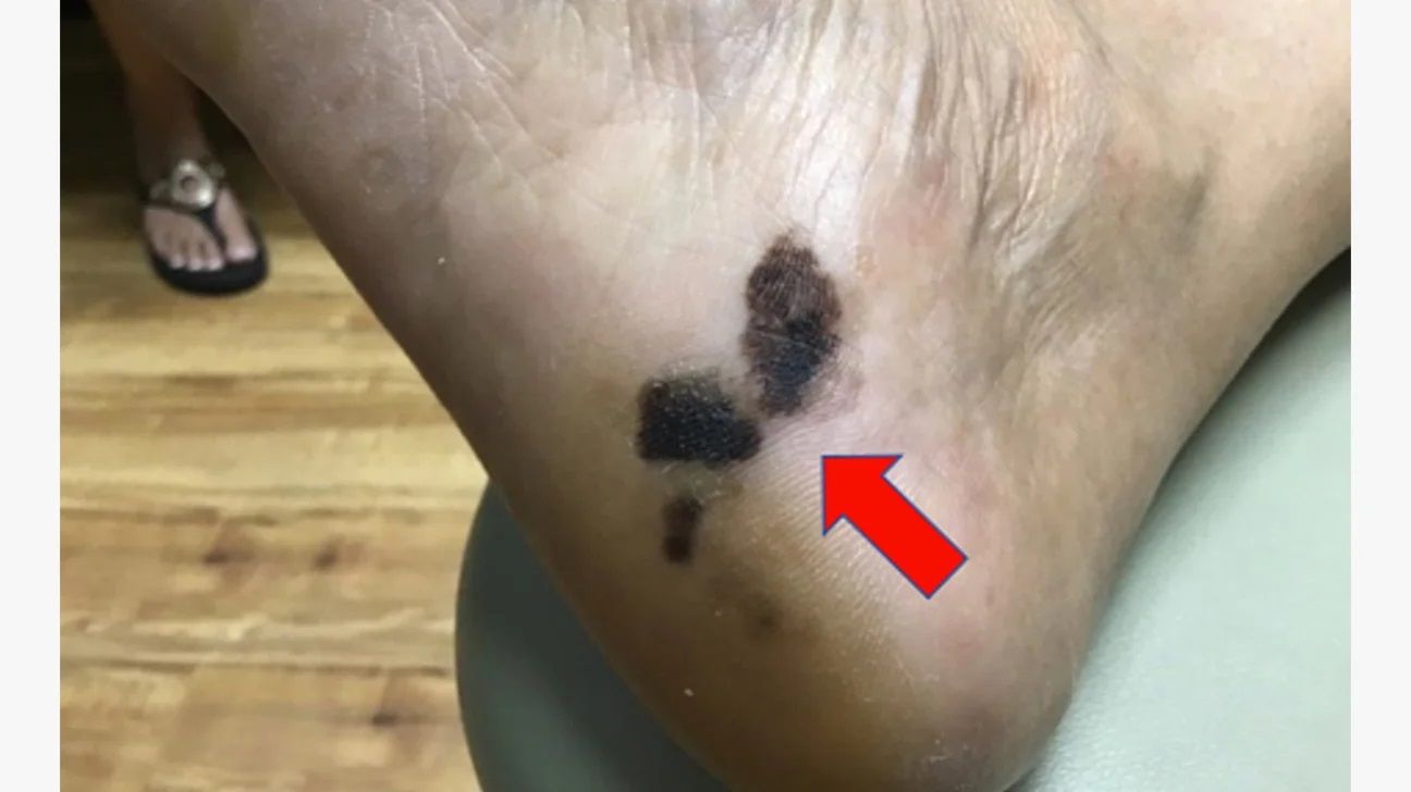
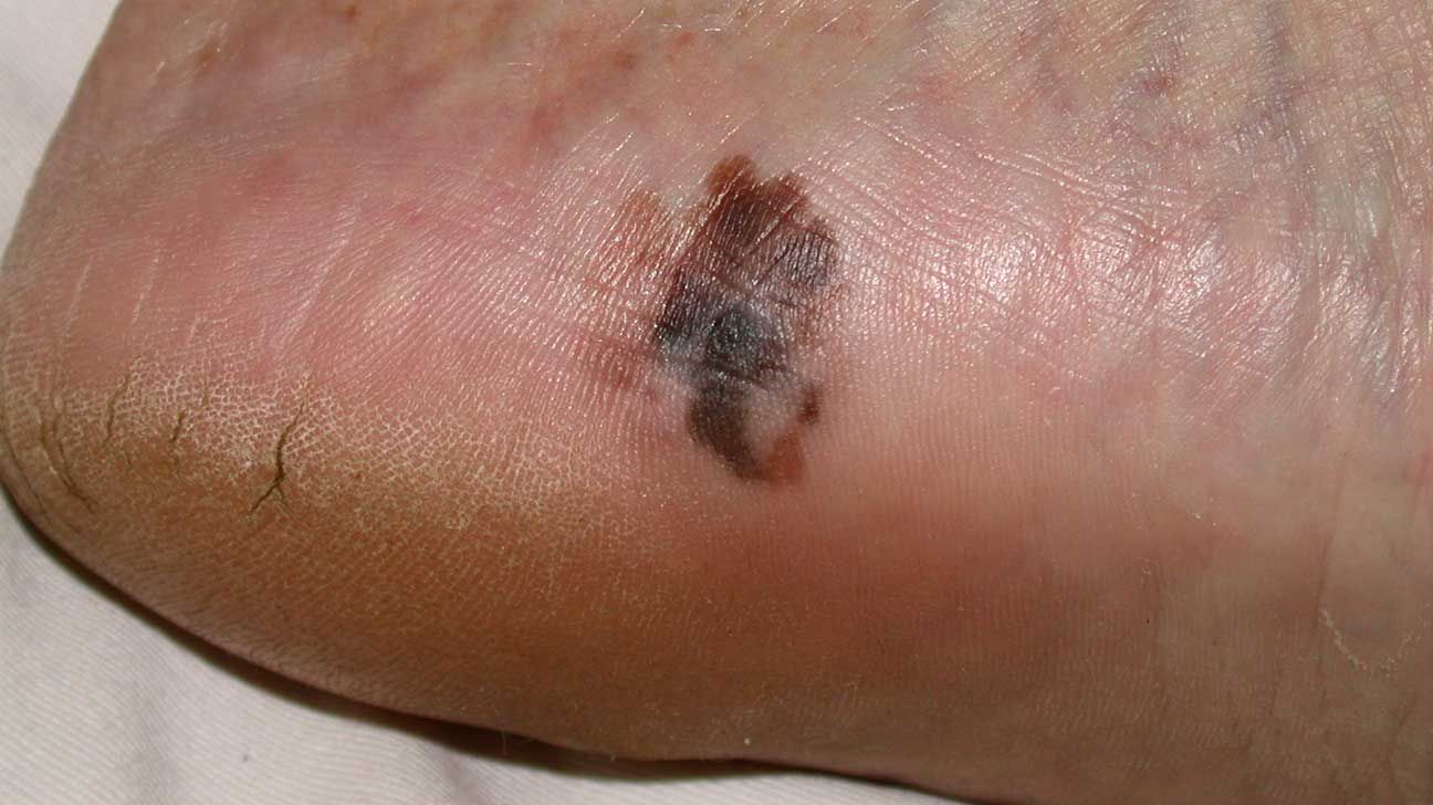
Acral lentiginous melanoma
Acral lentiginous melanoma is the most common form of malignant melanoma among individuals with darker skin tones. Warning signs include:
- a darkened or discolored patch, often on the palms, soles, fingers, toes, or beneath the nails, beginning as a slowly enlarging area
- a mark that resembles a bruise or stain
- location on the hands, feet, or nail beds
Rare types of melanoma
- Mucosal melanoma. A melanoma that develops in moist mucosal surfaces, including the eyes, mouth, vagina, and vulva.
- Desmoplastic melanoma. Typically found on chronically sun-damaged skin of the head and neck. It represents approximately 1 percent of melanomas in the United States.
- Uveal melanoma. A melanoma occurring in the eye that can lead to visual disturbances or loss. Early symptoms are uncommon and are often detected during routine eye exams. Later symptoms may include dark spots in vision, blurred sight, floaters, and visible changes in the eye’s shape or position.
Risk factors for melanoma
There are several factors that increase the likelihood of developing melanoma, including:
- frequent sunburns, especially blistering sunburns
- residing in areas with intense sunlight exposure
- use of tanning beds
- fair or freckled skin
- a personal or family history of melanoma
- having numerous moles
- a previous diagnosis of skin cancer
- a weakened immune system
Limiting ultraviolet (UV) exposure, wearing protective clothing, and applying broad-spectrum sunscreen are important preventive steps.
How are moles related to melanoma?
Nearly everyone has at least one mole — a flat or raised spot that may be pigmented or match the surrounding skin. Moles form when melanocytes cluster together.
They often appear during childhood, and by adulthood, many people have 10 or more moles.
Most moles remain benign and stable. However, some may enlarge, alter their shape, or change color. A small percentage can transform into melanoma, which is why comparing your skin to Skin cancer pictures early stages can be helpful for early recognition.
Look for changes in skin and moles
The most significant indicator that a skin lesion could be melanoma is change over time. A malignant mole typically evolves in size, shape, or color.
Dermatologists frequently recommend the ABCDE rule to evaluate suspicious moles:
- Asymmetry
- Border
- Color
- Diameter
- Evolving
Asymmetry
A symmetrical mole appears similar on both halves if divided down the center. In contrast, an asymmetrical mole has mismatched sides in size or contour. Cancer cells often grow unevenly and unpredictably.
Border
A typical mole has smooth, clearly defined edges. If the border looks blurred, ragged, or notched — as though the pigment extends beyond the lines — it may be concerning.
Color
Moles can normally be brown, black, or tan. However, multiple colors within a single lesion may indicate melanoma.
A melanoma may display varying shades of brown or black, or patches of white, red, gray, or blue. These color differences can appear differently depending on skin tone.
Diameter
Most benign moles measure about 6 millimeters (1/4 inch) or less, roughly the size of a pencil eraser.
Moles that exceed this size or continue to enlarge should be evaluated by a healthcare professional.
Evolving
Any mole that changes is a red flag. Perform routine skin self-exams and monitor for spots that grow, darken, or alter in texture.
In addition to the ABCDE criteria, watch for:
- redness
- scaling
- bleeding
- oozing
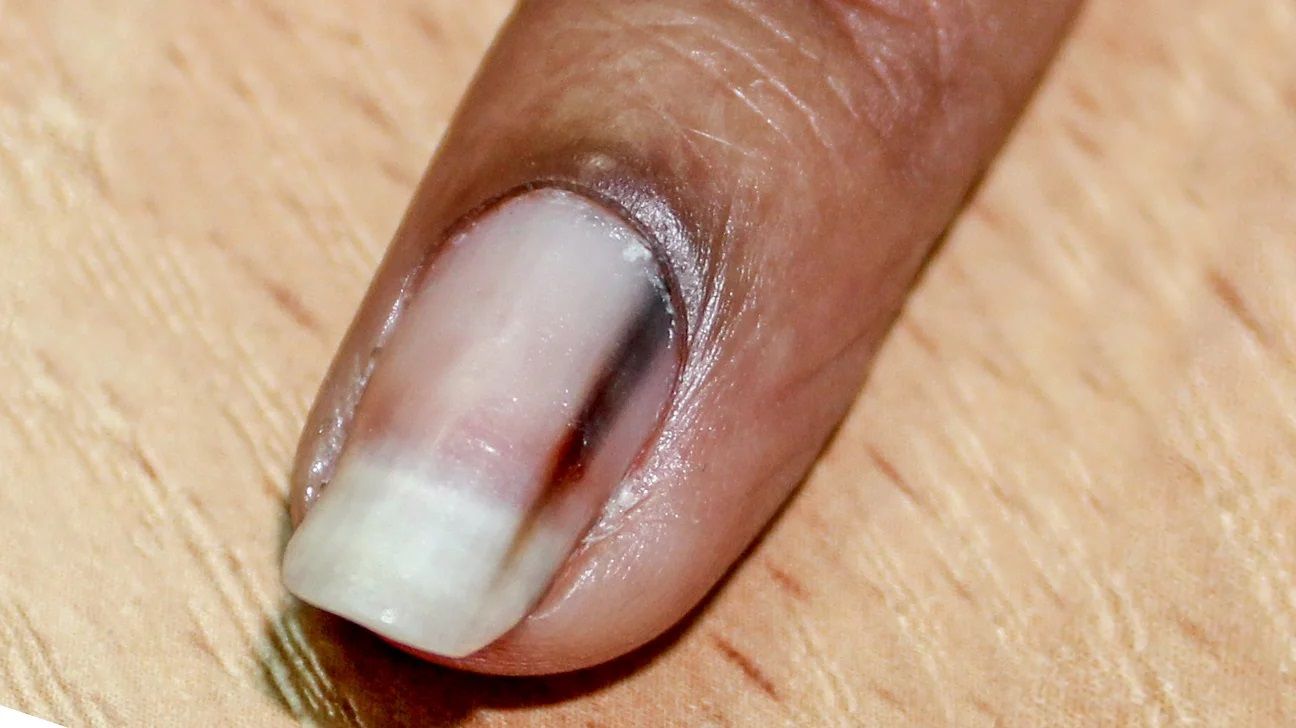
Nail melanoma
Although uncommon, melanoma can develop beneath the nails. This condition is known as subungual melanoma. It may present as a pigmented band running along the nail and can also:
- lead to thinning or splitting of the nail
- form nodules with bleeding
- widen toward the cuticle
Subungual melanoma does not always cause discomfort. Consult a doctor if you notice unusual nail discoloration or structural changes.
When to speak with a dermatologist
Performing regular skin checks increases the likelihood of identifying skin cancer at an early, treatable stage.
If you observe a new lesion or any unusual change, schedule an appointment with a dermatologist for a comprehensive skin examination.
Individuals with numerous moles or a family history of skin cancer should have routine dermatology visits. A dermatologist may create a mole map to track changes over time.
If a suspicious lesion is identified, a biopsy may be performed to test for cancer cells. When melanoma is detected, early removal is crucial to prevent spread.
Frequently asked questions
What does a melanoma look like when it first appears?
Melanoma often starts with visible alterations in an existing mole, including changes in size, shape, color, or texture. It may also present as a new, unusual-looking mole.
Is melanoma raised or flat?
Melanoma can appear either raised or flat, depending on the type. The four main types each have distinct visual features. A healthcare professional can confirm the diagnosis and subtype.
How can you tell if a spot is melanoma?
Any evolving mole or newly developed spot should be examined by a doctor. Only a qualified medical provider can determine whether a lesion is melanoma.
What are the three signs and symptoms of melanoma?
There are more than three signs of melanoma. Seek medical evaluation if you notice:
- an uneven shape
- irregular borders
- color variation or discoloration
- a size larger than a pencil eraser
- changes in size, contour, or color
- itching or fluid discharge
- a sore that fails to heal
- pigment spreading beyond the original spot
- redness or swelling extending to nearby skin
- pain, tenderness, or persistent itching
- a change in texture, such as scaliness, oozing, bleeding, or a new lump
Learn more about melanoma and how early detection can improve outcomes.


Leave a Reply
You must be logged in to post a comment.