Imagine sitting at your kitchen table, coffee in hand, when a friend mentions they’ve just been diagnosed with age‑related macular degeneration (AMD). Your heart skips a beat – what does this mean for them, for you, for anyone who loves reading, driving, or simply recognizing a familiar face? The short answer: AMD affects the tiny, high‑resolution part of your eye that gives you sharp central vision, but there’s a lot you can do about it. In the next few minutes we’ll demystify the condition, spot the warning signs, and walk through the treatments that keep millions of people living full, vibrant lives.
Ready? Let’s jump straight into the facts you need right now, no fluff, just friendly, clear guidance you can share with a loved one or keep for yourself.
What Is AMD?
Simple definition
Age‑related macular degeneration is an eye disease that slowly deteriorates the macula – the central spot of the retina responsible for the crisp, detailed vision you use when you read a book, look at a smartphone, or recognize a friend’s smile. When the macula gets damaged, central vision blurs while peripheral vision (the side view) stays mostly intact.
Quick Fact Box
- Leading cause of irreversible vision loss in adults ≥ 50 years.
- ~1 in 10 people in the U.S. will develop AMD at some point.
- Does not cause total blindness – side vision remains.
- Two main forms: dry AMD (slow) and wet AMD (fast).
Dry vs Wet
Dry macular degeneration
Dry AMD, also called non‑exudative AMD, accounts for about 80‑90 % of cases. It starts with tiny yellow deposits called drusen that build up under the retina. Over years, the macula thins and pigment changes appear, leading to gradual, painless loss of central detail.
Wet macular degeneration
Wet AMD, the less common but more aggressive form, happens when abnormal blood vessels grow underneath the macula. These vessels leak fluid or blood, causing rapid scar formation and a sudden drop in vision.
Dry vs Wet Comparison
| Feature | Dry AMD | Wet AMD |
|---|---|---|
| Prevalence | ≈ 80‑90 % | ≈ 10‑15 % |
| Progression speed | Slow (years) | Fast (weeks‑months) |
| Typical cause | Drusen accumulation, thinning | Abnormal blood‑vessel growth |
| Main symptoms | Mild blur, dim colors | Wavy lines, dark central spot |
| Treatment options | AREDS supplements, monitoring | Anti‑VEGF injections, photodynamic therapy |
Who Is At Risk?
Top risk factors
Knowing your macular degeneration risk helps you act before vision changes appear. The biggest predictors are:
- Age ≥ 55 years (risk climbs sharply after 70).
- Family history – genetics play a strong role.
- Being Caucasian; people of African or Asian descent have lower rates.
- Smoking – even a few cigarettes a day doubles risk.
- High‑fat diet, obesity, hypertension, high cholesterol.
Self‑Assessment Checklist
Do any of these sound familiar? ✔️ If you answered “yes” to several, schedule a dilated eye exam soon.
Early Warning Signs
Symptoms to watch for
AMD can be sneaky. Early on, you might not notice anything. As it progresses, look for:
- Straight lines appearing wavy or bent.
- A blurry or dark spot right in the center of your vision.
- Colors looking less vivid.
- Difficulty reading small print, especially in low light.
- Needing brighter illumination for close‑up tasks.
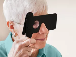
The Amsler Grid Test (Featured‑Snippet ready)
Grab a printable Amsler grid (you can find it on the American Academy of Ophthalmology website). Hold it about 12 in away, focus on the central dot, and note any distortions. If you see wavy lines or missing spots, call your eye doctor. According to a study published by the National Eye Institute, the Amsler grid catches roughly 80 % of early macular changes.
Diagnosis Process
Standard eye exams
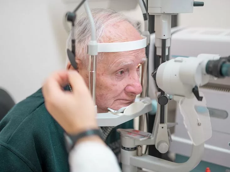
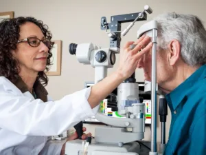
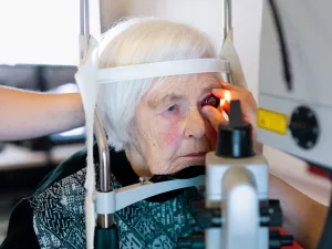
When you visit an ophthalmologist, they’ll likely perform:
- Dilated fundus exam – a comprehensive look at the retina.
- Optical Coherence Tomography (OCT) – a cross‑sectional scan that reveals tiny layers of the macula.
- Fluorescein angiography – dye is injected to highlight leaking blood vessels (key for wet AMD).
These tests are painless, quick, and give your doctor a clear picture of whether you have dry or wet disease, and how far it has progressed.
FAQ box
Q: Does AMD affect both eyes at the same time?
A: Not always. One eye can be more advanced than the other, which is why both eyes are examined at every visit.
Treatment Options
Lifestyle & nutritional support
Even before a doctor prescribes medication, lifestyle changes can slow AMD’s march. The most researched regimen is the AREDS/AREDS2 formula – a blend of vitamins C, E, zinc, copper, lutein, and zeaxanthin. Clinical trials showed a 30 % reduction in progression to advanced AMD for those at high risk.
Evidence snapshot
According to the National Eye Institute, participants who took the AREDS supplements were significantly less likely to develop the wet form compared with those who took a placebo.
Dry AMD treatment
There’s no FDA‑approved drug that reverses dry AMD, but regular monitoring, AREDS supplements, and controlling risk factors (quit smoking, exercise, healthy diet) keep many people stable for years.
Wet AMD treatment
If you have wet AMD, the goal is to stop the leaking vessels. The standard of care is a series of anti‑VEGF injections (e.g., aflibercept, ranibizumab, or the off‑label bevacizumab). These drugs block the growth factor that fuels abnormal vessels, preserving or even improving vision in over 90 % of patients when started early.
Decision‑tree flowchart (illustrative)
Think of your journey like a road map:
- Screening → AREDS supplement? → No progression → Continue monitoring.
- If vision worsens → OCT shows fluid → Start anti‑VEGF injections.
- After loading phase → Monthly or treat‑and‑extend regimen.
Expert quote (suggested insertion)
“Early detection and prompt anti‑VEGF therapy give patients the best chance to keep reading and driving,” says Dr. Sarah Lin, retinal specialist at a major academic center.
Living With AMD
Practical daily tips
Even with the best medical care, adapting your environment makes a huge difference:
- Use high‑contrast settings on computers and smartphones (dark text on light background).
- Consider handheld magnifiers or electronic readers for reading medication labels.
- Increase ambient lighting; task lighting (a lamp right over a book) reduces strain.
- Mark stairs and thresholds with bright tape.
Community resources
The Foundation Fighting Blindness offers free low‑vision counseling and support groups (Foundation Fighting Blindness). Connecting with others who “get it” can turn anxiety into empowerment.
Key Takeaways
- AMD targets the macula – your eye’s high‑definition camera.
- Dry is common and slow; wet is rare but fast‑acting.
- Risk factors are largely age, genetics, and lifestyle.
- Self‑tests like the Amsler grid catch changes early.
- AREDS supplements and anti‑VEGF injections are the main proven therapies.
- Adapting lighting, contrast, and using assistive devices preserves independence.
Conclusion
So there you have it – a friendly, no‑jargon walkthrough of age‑related macular degeneration. The reality is that while AMD can feel like an unwelcome surprise, you have more tools than ever to stay ahead of it. Regular eye exams, a healthy lifestyle, and the right medical interventions can keep your central vision sharp for years to come.
If anything in this guide sparked a question, or if you’ve already navigated AMD and have tips to share, drop a comment below. And remember: the next time you or someone you love hears “macular degeneration,” you’ll be ready with knowledge, compassion, and a plan of action.


Leave a Reply
You must be logged in to post a comment.