Heart valve testing is the series of imaging and functional exams that tell us whether a valve is leaking, narrowed, or working just fine. Knowing the answer right now can shape the next steps—whether it’s medication, a repair, or a brand‑new replacement heart valve.
If you or a loved one are facing a valve issue, the info below will walk you through why the tests matter, what each test does, and how doctors turn numbers into decisions. Think of it as a friendly chat over coffee, with a dash of science and a sprinkle of stories.
Why Testing Matters
What the tests reveal (benefits)
Every heartbeat is a tiny report card for your valves. The tests give us:
- Early detection of stenosis (narrowing) or regurgitation (leakage) before symptoms flare up.
- Clear guidance on whether medication, a repair, or a replacement heart valve is the best route.
- A roadmap for surgeons to plan the safest procedure, especially when child cardiac surgery is on the table.
Possible downsides (risks)
Like any medical step, there are a few things to keep in mind:
- False‑positive or false‑negative results can cause unnecessary worry or missed disease.
- Radiation exposure from CT or fluoroscopy—still low, but we aim for “as low as reasonably achievable.”
- Contrast agents may irritate kidneys; doctors’ll check your labs first.
Core Test Types
| Test | Primary Goal | Typical Setting | Key Metric |
|---|---|---|---|
| Transthoracic Echo (TTE) | Visualize valve motion & blood flow | Outpatient clinic | Ejection fraction, regurgitant jet area |
| Transesophageal Echo (TEE) | Higher‑resolution images of posterior valves | Hospital/OR | Leaflet morphology, clot detection |
| Cardiac MRI | 3‑D anatomy + flow quantification | Specialized imaging center | Velocity‑encoded phase contrast |
| CT Angiography | Calcification & annular sizing (TAVR planning) | Radiology suite | Agatston calcium score |
| Cardiac Catheterization | Direct pressure gradients, coronary anatomy | Cath lab | Peak‑to‑peak pressure gradient |
How each test works (plain language)
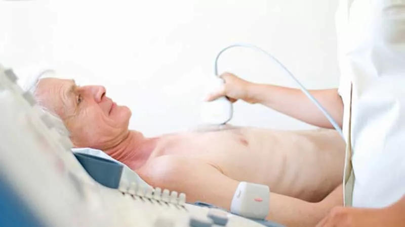
Echocardiogram (Echo) – Think of a sonar for your heart. High‑frequency sound waves bounce off the moving structures and create a moving picture on screen. The snippet you might have seen says an echo “uses high‑frequency sound waves (ultrasound) to make pictures of your heart.”
MRI – A giant magnet and radio waves spin the hydrogen atoms in your blood, letting us see flow patterns without any radiation. It’s great for patients who want to skip the X‑rays.
CT – X‑ray slices are compiled into a 3‑D model. It tells us exactly how much calcium is built up on the leaflets, which is crucial when sizing a replacement heart valve for a transcatheter procedure.
TEE – A tiny probe slides down the throat, getting close to the heart for crystal‑clear images—especially useful for spotting small clots or subtle leaflet problems.
Catheterization – A thin tube slides into a blood vessel and measures pressure directly on each side of the valve. It’s the “gold standard” for confirming the severity of a stenosis.
Industry Insight
When it comes to durability testing of prosthetic valves, labs use accelerated wear testing (AWT). According to ViVitro Labs’ Dual Control Technology, modern AWT keeps leaflet closing velocity constant, avoiding unrealistic stress that could mislead design decisions. Similarly, BDC Labs’ VDT‑3600i system offers six independent stations, letting engineers test multiple valve sizes at once.
Special Patient Groups
Pediatric heart‑valve testing
Kids aren’t just small adults; their hearts beat faster and their anatomy is tinier. Pediatric cardiology teams often start with a quick transthoracic echo, which can capture congenital heart defects in seconds. When a more detailed look is needed, they may use a child‑size TEE under mild sedation. The goal is to see the problem without exposing a child to radiation whenever possible.
Read more about the unique world of pediatric cardiology and why it matters.
Adults with degenerative disease
As we age, calcium can build up on valve leaflets. A CT scan often becomes the star of the show, highlighting the exact amount of calcification and helping the surgeon select the right size for a replacement heart valve. Echo still tells us whether the valve is leaking, but CT gives the three‑dimensional roadmap surgeons need.
High‑risk or frail patients
For patients who can’t tolerate long procedures, doctors may rely heavily on echo and MRI, skipping the radiation‑heavy CT unless absolutely necessary. The focus shifts to “least invasive, most information.”
Reading The Results
Normal vs. abnormal thresholds (quick reference)
- Mean pressure gradient > 20 mmHg generally indicates moderate stenosis.
- Regurgitant volume > 60 ml suggests severe leakage.
- Effective orifice area (EOA) < 1.0 cm² for an aortic valve is considered severe.
What the cardiology team does next
Based on the numbers, three paths usually appear:
- Watchful waiting – If the disease is mild and you feel fine, doctors may schedule a repeat echo in 1‑2 years.
- Medical therapy – Blood‑thinners, ACE‑inhibitors, lifestyle tweaks (low‑salt diet, regular exercise).
- Intervention – Valve repair, minimally invasive TAVR (transcatheter aortic valve replacement), or open‑heart surgery with a new prosthetic valve.
Real World Stories
Case 1: A 3‑month‑old baby presented with a bicuspid aortic valve that was already showing signs of narrowing. A quick TTE caught the problem, followed by a low‑dose CT to gauge calcium. The infant underwent a balloon valvuloplasty and later required a planned pediatric heart valve surgery.
Case 2: 58‑year‑old teacher felt occasional shortness of breath. A TEE revealed moderate mitral regurgitation. After medication didn’t fully help, the doctor recommended a percutaneous mitral‑clip—an outpatient repair that avoided open surgery.
Case 3: 72‑year‑old retiree had heavy calcium on her aortic valve. CT showed a calcium score of 850 AU. The heart team chose a transcatheter replacement heart valve (TAVR) and she walked out the same day.
Future Of Testing
3‑D printed models
Surgeons now print patient‑specific valve replicas from CT data. Holding a tangible model helps them rehearse the operation, reducing surprises in the OR.
AI‑assisted echo interpretation
Machine‑learning algorithms can automatically measure gradients and regurgitant volumes, offering faster, more consistent readings. A recent study in the Journal of Cardiac Imaging showed AI reduced interpretation time by 40 % without sacrificing accuracy.
Next‑gen durability labs
Advanced AWT machines are able to simulate years of wear in just a few weeks, giving manufacturers confidence that a new prosthetic will survive a lifetime. The combination of better simulation and real‑world data is raising the bar for safety.
How To Prepare For Your Appointment
Checklist for patients
- Gather any previous imaging reports (echo, CT, MRI).
- Write down current medications—especially blood thinners.
- If you have a scheduled TEE, follow fasting instructions (usually nothing solid for 6 hours).
- Bring a list of questions you want to ask the cardiologist.
Questions to ask your doctor
- “What does my pressure gradient tell us about urgency?”
- “Are there less‑invasive options for my valve type?”
- “How will the test results influence my long‑term care plan?”
Conclusion
Heart valve testing gives a clear picture of how well your valve works, helping doctors weigh the benefits of monitoring, medication, repair, or a replacement heart valve. By understanding what each test does, its risks, and how results guide care—for both adults and children—you can ask the right questions and feel confident about the next steps. If you’re scheduled for any of these exams, bring your reports, jot down your concerns, and remember that the whole team is there to keep your heart ticking smoothly.


Leave a Reply
You must be logged in to post a comment.