Mouth cancer pictures early stages often show subtle white patches, red patches, or sores on the tongue that may not cause discomfort at first. Unlike many harmless mouth lesions, these early abnormalities are typically painless when they initially develop, which can make them easy to overlook.

Oral cancer is a type of cancer that develops in the tissues of the mouth or throat. It may involve any of the functional structures within your oral cavity, including the:
- lips
- lining of the lips and cheeks
- front two-thirds of the tongue (the back third of the tongue, or base, is considered part of the oropharynx, or throat)
- gums
- floor of the mouth (under the tongue)
- roof of the mouth
While teeth themselves cannot develop cancer, cancer in the surrounding tissues can affect nearby teeth and supporting structures.
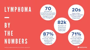
The American Cancer Society (ACS) estimates that 59,660 people will be diagnosed with oral cavity cancer or oropharyngeal cancer in 2025, and 12,770 of those cases will result in death.
Reviewing Mouth cancer pictures early stages and understanding the warning signs can help you recognize suspicious changes promptly. Below, you’ll learn what oral cancer may look and feel like, how it differs from common mouth sores, and when to consult a healthcare professional.
Pictures of oral cancer
What does cancer of the mouth look like?
The surfaces of your mouth, lips, and tongue are covered by flat cells known as squamous cells. According to the ACS, the majority of mouth cancers originate in these cells.
An unexplained white or red patch on your tongue, gums, tonsils, or inner cheek lining may be an early indication of squamous cell carcinoma. In many Mouth cancer pictures early stages, these areas appear flat at first but may gradually thicken or change texture.
The appearance and sensation of oral cancer can differ from person to person. The affected area may feel firm, thickened, or slightly raised. Some people develop a persistent ulcer, erosion, or roughened patch that does not heal.

One key factor is persistence. Noncancerous mouth lesions often improve within a few weeks. If you notice a red or white patch that remains for more than 2 weeks, it’s important to see a dentist or doctor for evaluation.
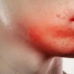
Red patches
Bright red, velvety-looking areas inside the mouth are called erythroplakia. These lesions are frequently considered precancerous, meaning they have the potential to become cancerous.
A 2022 review found that approximately 1 in 5 cases of erythroplakia progress to cancer, a lower percentage than earlier research suggested.
Even so, any vivid red lesion in the mouth warrants attention. If erythroplakia is suspected, a dentist may perform a biopsy. This can involve removing a thin layer of tissue or collecting cells using a specialized brush for laboratory analysis.
White patches
A white or gray patch inside the mouth or on the lips is known as leukoplakia. These areas may feel thick, rough, or hardened and are often difficult to scrape away.
Cell changes leading to leukoplakia may be triggered by include:
- chronic irritation from a rough tooth, broken denture, or tobacco use
- habitual chewing of the inner cheeks or lips
- exposure to carcinogenic substances
Although leukoplakia indicates abnormal tissue growth and carries a risk of malignancy, in most cases it is benign.
These patches usually form gradually over weeks or months. Comparing changes with reliable Mouth cancer pictures early stages can help you determine whether a professional exam is needed. For visual references involving gum involvement, you may also review Gum cancer pictures.
Mixed red and white patches
When red and white areas appear together, the condition is called erythroleukoplakia. This mixed lesion represents abnormal cell growth and carries a higher likelihood of becoming cancerous than leukoplakia alone. Often, these patches are visible before they cause any discomfort.
In early-stage disease, pain is uncommon. That’s why routine self-checks and dental visits are essential for early detection.
How to detect mouth cancer at home?
Erythroplakia can develop anywhere in the oral cavity, but it most often appears on the inner cheeks or the floor of the mouth.
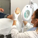
Perform a careful self-exam once a month in a well-lit room. Use a magnifying mirror to inspect all areas clearly.

With clean fingers, gently extend your tongue and check underneath. Examine both sides of the tongue, the inside of your cheeks, and the inner and outer surfaces of your lips. Pay close attention to dark spots, persistent ulcers, or unusual discoloration. For example, reviewing Oral cancer black spot on gums photos may help you identify concerning pigment changes on the gums.
Be alert for lumps, thickened areas, unexplained bleeding, or sores that fail to heal. Early recognition significantly improves treatment outcomes.
Canker sores vs. oral cancer
A canker sore typically causes burning, stinging, or tingling before it becomes visible.
By contrast, mouth cancer in its early stages seldom causes pain. Abnormal cell growth often appears as flat, discolored patches rather than tender ulcers.
Canker sores usually have a shallow, crater-like center. The middle may look white, gray, or yellow, surrounded by a red border.
Although canker sores can be uncomfortable, they are not malignant, meaning they do not turn into cancer.
Most canker sores heal within 2 weeks. Any sore, lump, or patch that persists beyond that timeframe should be examined by a healthcare professional.
When to seek medical help
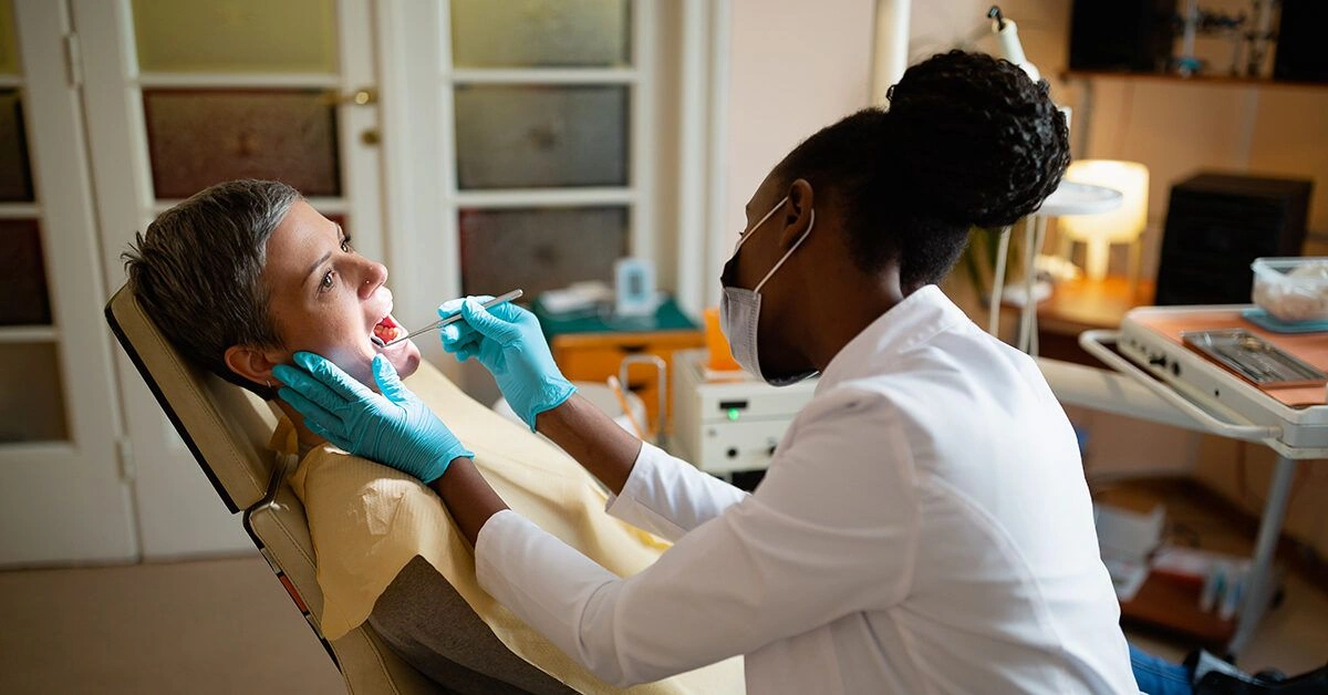
If you think you may have signs of oral cancer, schedule an appointment with a dentist or doctor. The Society of Behavioral Medicine states that trained clinicians can conduct a visual examination to identify suspicious lesions.
You’ll also be asked about symptoms. Additional signs of mouth cancer may include:
- persistent mouth pain
- trouble swallowing
- difficulty speaking
- unexplained weight loss
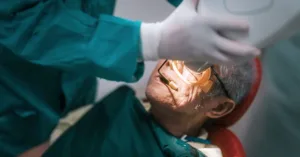
If an abnormal area is found, a biopsy may be performed to determine whether cancerous cells are present. Early diagnosis greatly increases the likelihood of successful treatment.
Can regular screenings help with prevention?
Routine dental checkups twice a year serve as an important screening measure. Dentists are trained to detect subtle changes that may indicate early oral cancer. Physicians may also examine the mouth during standard physical exams.
Addressing precancerous changes promptly reduces the chance that abnormal cells will progress to malignancy. Reviewing Mouth cancer pictures early stages and staying informed can empower you to seek care quickly.
You can further lower your risk by avoiding tobacco products, including “dip,” “chew,” and cigarettes. Research consistently links tobacco use to an increased risk of mouth cancer. Limiting alcohol intake and maintaining good oral hygiene may also support overall oral health.
The bottom line
Most mouth sores are not cancerous and resolve on their own.
However, any white, red, or mixed patch in your mouth or on your lips that does not improve within a few weeks should be evaluated by a dentist or doctor.
A healthcare professional can determine whether a lesion is cancerous or noncancerous and guide you on appropriate next steps. Early detection remains one of the most effective tools in improving outcomes for oral cancer.


Leave a Reply
You must be logged in to post a comment.