A shoulder computed tomography scan (CT or CAT scan) produces cross-sectional pictures of the shoulder by using advanced X-ray equipment. This imaging test enables physicians to visualize the shoulder’s bones and soft tissues to uncover irregularities. It can also assist in detecting tumors and clots.

A CT scan may be done with or without contrast dye. The contrast agent helps clinicians evaluate important blood vessels and anatomical structures and can reveal abnormalities that are otherwise hidden.
What is the purpose of a shoulder CT scan?
The primary reason for ordering a shoulder CT scan is to examine the shoulder after an injury. This could involve a single traumatic event or repeated problems, such as a shoulder that frequently slips out of its socket or dislocates. The scan can provide a clearer view of a fracture or confirm a suspected break.
Your physician might request a shoulder CT scan to:
- detect blood clots
- locate masses or tumors
- identify infections
- reveal tears in muscles, tendons, or ligaments
- spot joint inflammation
- evaluate trauma-related injuries such as dislocations or fractures
- assist in surgical planning
- guide treatment decisions for your injury
In some cases, a shoulder CT scan is ordered to investigate joint complaints like pain, stiffness, or grinding noises — particularly when an MRI is not feasible (for example, for people with cardiac pacemakers). It can also help assess conditions related to a lump on shoulder if imaging detail of bone and soft tissue is needed.
What are the risks of a shoulder CT scan?
A shoulder CT scan carries minimal risk.
The contrast dye may provoke an allergic response or affect kidney function. This risk increases if you already have compromised kidney function from disease or infection. Modern contrast agents are generally much safer for the kidneys than older formulations.
As with any X-ray procedure, there is exposure to ionizing radiation during a CT scan. The radiation doses used are considered safe for adults but are not recommended for a developing fetus. Inform your physician if you are pregnant or suspect you might be.
How should I prepare for a shoulder CT scan?
Because the exam is noninvasive, preparation is usually simple.
Wear loose, comfortable clothing since you will lie down on an examination table. You will also be asked to remove jewelry and other metal items from your person.
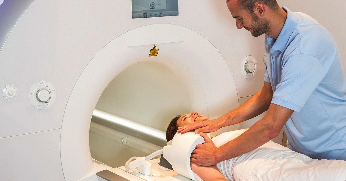
How is a shoulder CT scan performed?
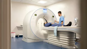
A CT scan is completed in the radiology department of a hospital or at a specialized imaging center. After removing jewelry and changing into a gown if required, a CT technologist will position you on the table.
If the scan uses contrast, an intravenous line will be placed — a small needle inserted into a vein in your arm so the dye can be injected. The discomfort is minimal, often described as similar to a standard blood draw.
Your technologist may instruct you to lie in a certain orientation and may use cushions or straps to keep you steady long enough to capture high-quality images. You may also be asked to hold your breath briefly during individual scans to avoid motion blur.
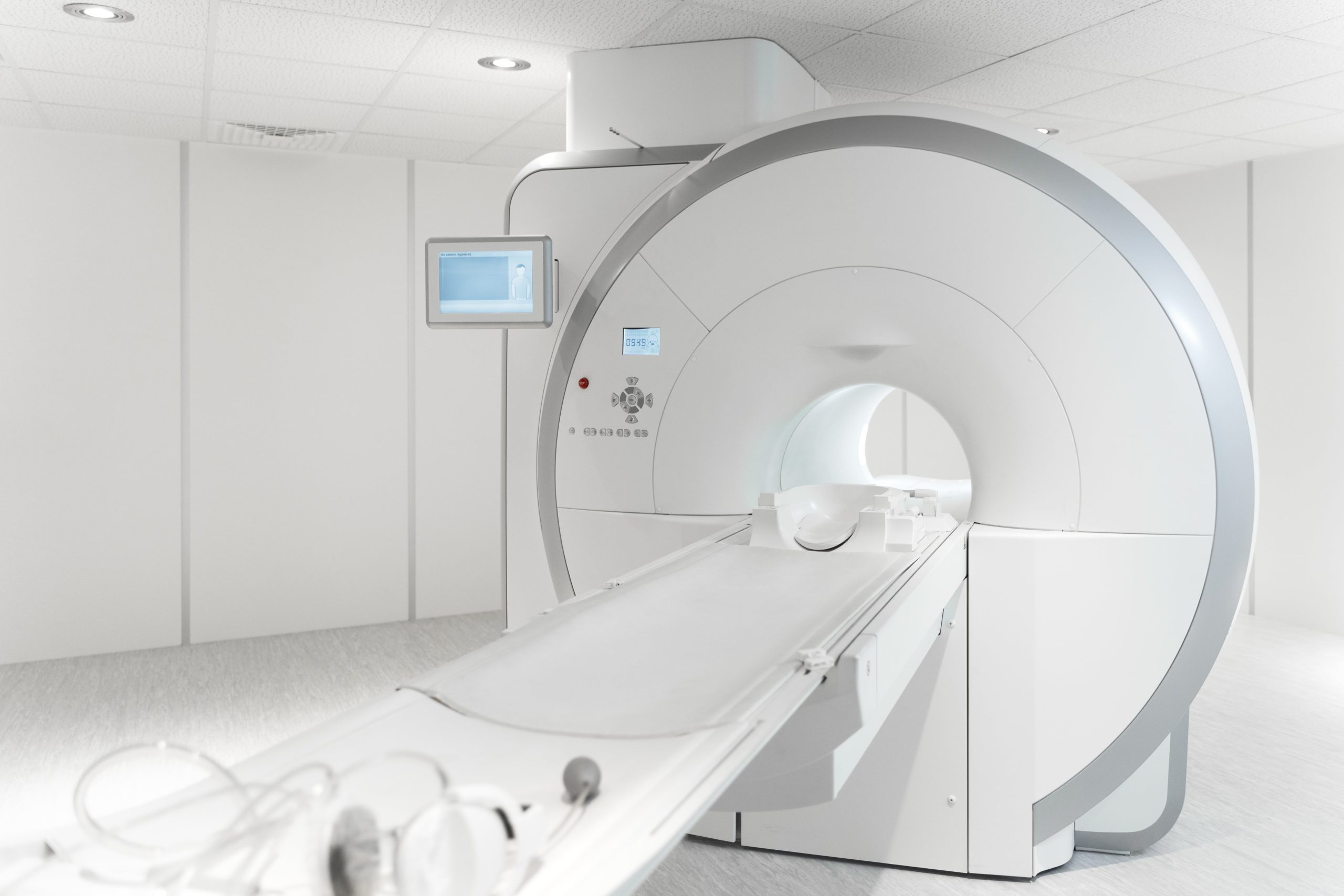
The technologist operates the scanner from an adjacent room, using controls to slide the table into the CT unit. The machine resembles a large ring of plastic and metal that rotates around you while the table moves through the opening.
After one series of images, you might wait while the technologist inspects the pictures to ensure they are sufficiently clear for interpretation.
When the scanning session is finished, you can change back into your own clothes and resume normal activities.
Most CT scans require roughly 30 to 45 minutes from start to finish.
After a shoulder CT scan
CT scan results usually take about a day to be processed. Your physician will arrange a follow-up visit to review the findings and recommend next steps based on the imaging results.


Leave a Reply
You must be logged in to post a comment.