Your first pregnancy ultrasound is often an eagerly anticipated milestone: around 12 or 13 weeks pregnant, you’re excited to catch a first glimpse of the little person who’s been developing for the last three months. Sure, they’ll likely look a bit alien at that stage — but they’ll be your alien, and you want to see them.
Sometimes an ultrasound is recommended much earlier, though, and we’ll be frank: it feels different. That’s because a great deal of embryonic development happens between weeks 7 and 12, so an early scan can be a very different experience from the typical first-trimester ultrasound.
A 7-week scan may not deliver the emotional bonding moment you’re hoping for, since many details may not yet be visible. Still, here’s what to expect.
Why your clinician might request an early ultrasound
While a 7-week ultrasound isn’t routine for everyone, there are a number of legitimate reasons your healthcare provider might want one — and not all of them are dire.
In fact, the most frequent reason for ordering an ultrasound before 12 or 13 weeks is to determine the pregnancy date more precisely.
If your symptoms don’t align with the date from your last period or there’s uncertainty about how far along you are, measurements from an early scan can give your clinician a clearer estimate of gestational age.
Other reasons for an early ultrasound include:
- Confirming multiples. If you’ve had fertility treatments or suspect more than one embryo, you may want confirmation early on.
- Confirming a fetal heartbeat. If you’ve experienced spotting or bleeding, your provider will check whether a miscarriage is occurring or investigate the cause of unexplained bleeding.
- Ruling out an ectopic pregnancy. When an embryo implants outside the uterus, pregnancy symptoms and a positive test can still occur even though the embryo isn’t viable. Untreated ectopic pregnancy can be life-threatening, so prompt diagnosis is essential.
- Evaluating reproductive anatomy. Problems with the uterus, cervix, ovaries, or fallopian tubes can complicate pregnancy. If your clinician suspects an issue — for example, uterine fibroids — they may want that information early.
So, a 7-week ultrasound isn’t necessarily a sign of trouble; it could simply be a way to gather important information to support a healthy pregnancy.
What happens at the appointment
Popular culture may have taught you that your first ultrasound involves a technician gliding a wand over your belly and unveiling a clear image of a tiny, peaceful baby floating in your uterus.
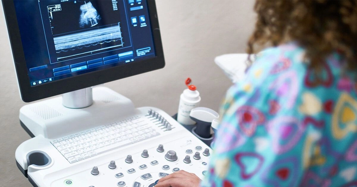
That’s typically not the case at 7 weeks, though. At this stage your embryo is often too small to be clearly visualized with a transabdominal scan. Instead, a transvaginal ultrasound is usually performed.
We won’t sugarcoat it: it’s less comfortable. A technician inserts an ultrasound probe, called a transducer, a few inches into the vagina until it reaches the area of the cervix.
The technician will hold and gently adjust the probe as needed to obtain clear images of the uterus. It’s not usually painful, but it can be uncomfortable.
The sensation is comparable to the pressure you might feel during a routine gynecologic exam. The scan can take a bit longer than an abdominal ultrasound, which may add to discomfort, but technicians are trained to make the experience as tolerable as possible.
The upside: the procedure poses no risk to your embryo and uses no radiation. So although it may not be a pleasant memory, it won’t harm your baby.

What you might see on the screen
You won’t be counting tiny fingers and toes at this appointment; the embryo is usually far too small for detailed features to be visible. You may perceive a general shape or simply see evidence that something is present, but it’s also normal if nothing distinctly baby-like is apparent.
One thing you’re likely to spot in a healthy early pregnancy is the fetal heartbeat. It may be as rapid as roughly 110 beats per minute or more. If the embryo is positioned well for imaging, you might notice a flicker or pulsing on the screen — and you may hear the heartbeat briefly.
Typical structures visible around 7 weeks include:
- Gestational sac. One of the earliest sonographic signs of pregnancy, this is the fluid-filled cavity surrounding the embryo. It usually appears by about 5 weeks’ gestation and, when visualized, typically confirms an intrauterine pregnancy. On the scan it appears as a dark, round or oval space set off against the brighter uterine tissue.
- Yolk sac. Often visible before the embryo itself, the yolk sac is the first structure to form inside the gestational sac and supplies nutrients and oxygen until the placenta takes over. It looks like a small white ring or bubble within the sac.
- Fetal pole. The fetal pole is the initial sign that an embryo is forming inside the gestational sac. It appears as a dense, whitish mass attached to the yolk sac and may be curved or elongated depending on exact dating. A fetal pole with cardiac activity can typically be seen by transvaginal ultrasound around 6 weeks.
Which measurements are taken — and why
Beyond checking for cardiac activity, the main goal of a 7-week ultrasound is to measure early pregnancy structures so your clinician can estimate how far along you are.
That’s why this is often called a dating scan: the dimensions are useful indicators of gestational age.
The sonographer will measure the gestational sac and, if visible, take a crown-rump length (CRL) of the embryo. At 7 weeks the embryo typically measures about 5 to 9 millimeters and the gestational sac is commonly around 18 to 24 millimeters.
Embryonic growth is rapid at this stage, with notable increases from one week to the next.
A gestational sac markedly smaller than 18 mm usually suggests you’re less advanced — for example, 5 or 6 weeks rather than 7 — while a sac much larger than 24 mm may indicate an older gestational age.
Remember that ultrasound isn’t perfectly precise: fetal position and other factors can affect how accurate measurements are or whether they can be obtained at all.
If images are inconclusive, your clinician may recommend repeating the ultrasound in one to two weeks.
What it might mean if nothing is visible
Because many people don’t suspect pregnancy until at least 3 or 4 weeks after conception and the gestational sac is the first structure to form, a healthy pregnancy at the time of a 7-week scan will often show at least that initial development.
Still, you might not yet see a yolk sac, fetal pole, embryo shape, or heartbeat. If that’s the case, try not to panic.
You could be earlier in pregnancy than you believed, perhaps due to later ovulation than estimated. A tilted uterus can also make visualization more challenging until the embryo grows larger.
However, a 7-week scan can also reveal complications.
If there are absent or inconsistent signs — for example, a large gestational sac without a yolk sac or fetal pole — it could indicate a blighted ovum or an early miscarriage. These outcomes are common in the earliest weeks when the risk of loss is higher.
If you’re still having pregnancy symptoms but no intrauterine fetal development is detected, the sonographer and clinician will likely search for evidence of an ectopic pregnancy, and may use blood tests and a pelvic exam as part of the evaluation.
Can twins be seen this early?
Yes — particularly fraternal twins. Determining the number of embryos is one of the primary reasons clinicians perform early scans.
With fraternal twins (two separate fertilized eggs
Your first pregnancy ultrasound is often an eagerly anticipated milestone: around 12 or 13 weeks pregnant, you’re excited to catch a first glimpse of the little person who’s been developing for the last three months. Sure, they’ll likely look a bit alien at that stage — but they’ll be your alien, and you want to see them.
Sometimes an ultrasound is recommended much earlier, though, and we’ll be frank: it feels different. That’s because a great deal of embryonic development happens between weeks 7 and 12, so an early scan can be a very different experience from the typical first-trimester ultrasound.
A 7-week scan may not deliver the emotional bonding moment you’re hoping for, since many details may not yet be visible. Still, here’s what to expect.
Why your clinician might request an early ultrasound
While a 7-week ultrasound isn’t routine for everyone, there are a number of legitimate reasons your healthcare provider might want one — and not all of them are dire.
In fact, the most frequent reason for ordering an ultrasound before 12 or 13 weeks is to determine the pregnancy date more precisely.
If your symptoms don’t align with the date from your last period or there’s uncertainty about how far along you are, measurements from an early scan can give your clinician a clearer estimate of gestational age.
Other reasons for an early ultrasound include:
- Confirming multiples. If you’ve had fertility treatments or suspect more than one embryo, you may want confirmation early on.
- Confirming a fetal heartbeat. If you’ve experienced spotting or bleeding, your provider will check whether a miscarriage is occurring or investigate the cause of unexplained bleeding.
- Ruling out an ectopic pregnancy. When an embryo implants outside the uterus, pregnancy symptoms and a positive test can still occur even though the embryo isn’t viable. Untreated ectopic pregnancy can be life-threatening, so prompt diagnosis is essential.
- Evaluating reproductive anatomy. Problems with the uterus, cervix, ovaries, or fallopian tubes can complicate pregnancy. If your clinician suspects an issue — for example, uterine fibroids — they may want that information early.
So, a 7-week ultrasound isn’t necessarily a sign of trouble; it could simply be a way to gather important information to support a healthy pregnancy.
What happens at the appointment
Popular culture may have taught you that your first ultrasound involves a technician gliding a wand over your belly and unveiling a clear image of a tiny, peaceful baby floating in your uterus.
That’s typically not the case at 7 weeks, though. At this stage your embryo is often too small to be clearly visualized with a transabdominal scan. Instead, a transvaginal ultrasound is usually performed.
We won’t sugarcoat it: it’s less comfortable. A technician inserts an ultrasound probe, called a transducer, a few inches into the vagina until it reaches the area of the cervix.
The technician will hold and gently adjust the probe as needed to obtain clear images of the uterus. It’s not usually painful, but it can be uncomfortable.
The sensation is comparable to the pressure you might feel during a routine gynecologic exam. The scan can take a bit longer than an abdominal ultrasound, which may add to discomfort, but technicians are trained to make the experience as tolerable as possible.
The upside: the procedure poses no risk to your embryo and uses no radiation. So although it may not be a pleasant memory, it won’t harm your baby.
What you might see on the screen
You won’t be counting tiny fingers and toes at this appointment; the embryo is usually far too small for detailed features to be visible. You may perceive a general shape or simply see evidence that something is present, but it’s also normal if nothing distinctly baby-like is apparent.
One thing you’re likely to spot in a healthy early pregnancy is the fetal heartbeat. It may be as rapid as roughly 110 beats per minute or more. If the embryo is positioned well for imaging, you might notice a flicker or pulsing on the screen — and you may hear the heartbeat briefly.
Typical structures visible around 7 weeks include:
- Gestational sac. One of the earliest sonographic signs of pregnancy, this is the fluid-filled cavity surrounding the embryo. It usually appears by about 5 weeks’ gestation and, when visualized, typically confirms an intrauterine pregnancy. On the scan it appears as a dark, round or oval space set off against the brighter uterine tissue.
- Yolk sac. Often visible before the embryo itself, the yolk sac is the first structure to form inside the gestational sac and supplies nutrients and oxygen until the placenta takes over. It looks like a small white ring or bubble within the sac.
- Fetal pole. The fetal pole is the initial sign that an embryo is forming inside the gestational sac. It appears as a dense, whitish mass attached to the yolk sac and may be curved or elongated depending on exact dating. A fetal pole with cardiac activity can typically be seen by transvaginal ultrasound around 6 weeks.
Which measurements are taken — and why
Beyond checking for cardiac activity, the main goal of a 7-week ultrasound is to measure early pregnancy structures so your clinician can estimate how far along you are.
That’s why this is often called a dating scan: the dimensions are useful indicators of gestational age.
The sonographer will measure the gestational sac and, if visible, take a crown-rump length (CRL) of the embryo. At 7 weeks the embryo typically measures about 5 to 9 millimeters and the gestational sac is commonly around 18 to 24 millimeters.
Embryonic growth is rapid at this stage, with notable increases from one week to the next.
A gestational sac markedly smaller than 18 mm usually suggests you’re less advanced — for example, 5 or 6 weeks rather than 7 — while a sac much larger than 24 mm may indicate an older gestational age.
Remember that ultrasound isn’t perfectly precise: fetal position and other factors can affect how accurate measurements are or whether they can be obtained at all.
If images are inconclusive, your clinician may recommend repeating the ultrasound in one to two weeks.
What it might mean if nothing is visible
Because many people don’t suspect pregnancy until at least 3 or 4 weeks after conception and the gestational sac is the first structure to form, a healthy pregnancy at the time of a 7-week scan will often show at least that initial development.
Still, you might not yet see a yolk sac, fetal pole, embryo shape, or heartbeat. If that’s the case, try not to panic.
You could be earlier in pregnancy than you believed, perhaps due to later ovulation than estimated. A tilted uterus can also make visualization more challenging until the embryo grows larger.
However, a 7-week scan can also reveal complications.
If there are absent or inconsistent signs — for example, a large gestational sac without a yolk sac or fetal pole — it could indicate a blighted ovum or an early miscarriage. These outcomes are common in the earliest weeks when the risk of loss is higher.
If you’re still having pregnancy symptoms but no intrauterine fetal development is detected, the sonographer and clinician will likely search for evidence of an ectopic pregnancy, and may use blood tests and a pelvic exam as part of the evaluation.
Can twins be seen this early?
Yes — particularly fraternal twins. Determining the number of embryos is one of the primary reasons clinicians perform early scans.
With fraternal twins (two separate fertilized eggs), each baby typically has its own gestational sac. If your dating is correct, multiple sacs are often visible on a transvaginal ultrasound at 7 weeks.
With identical twins (one fertilized egg that split), there may be a single gestational sac but multiple yolk sacs, fetal poles, and heartbeats visible.
Bear in mind that ultrasounds are not infallible. You may not be far enough along for all these features to be detected yet, and embryos can sometimes hide behind each other. Multiple sacs or embryos may become clearer on a later scan.
Bottom line
Try not to be alarmed if your provider recommends a 7-week ultrasound; there are many valid reasons for this early check. It’s an uncomfortable but safe procedure that yields valuable information about whether a pregnancy is viable, how far along it is, and whether more than one embryo is present.


Leave a Reply
You must be logged in to post a comment.