
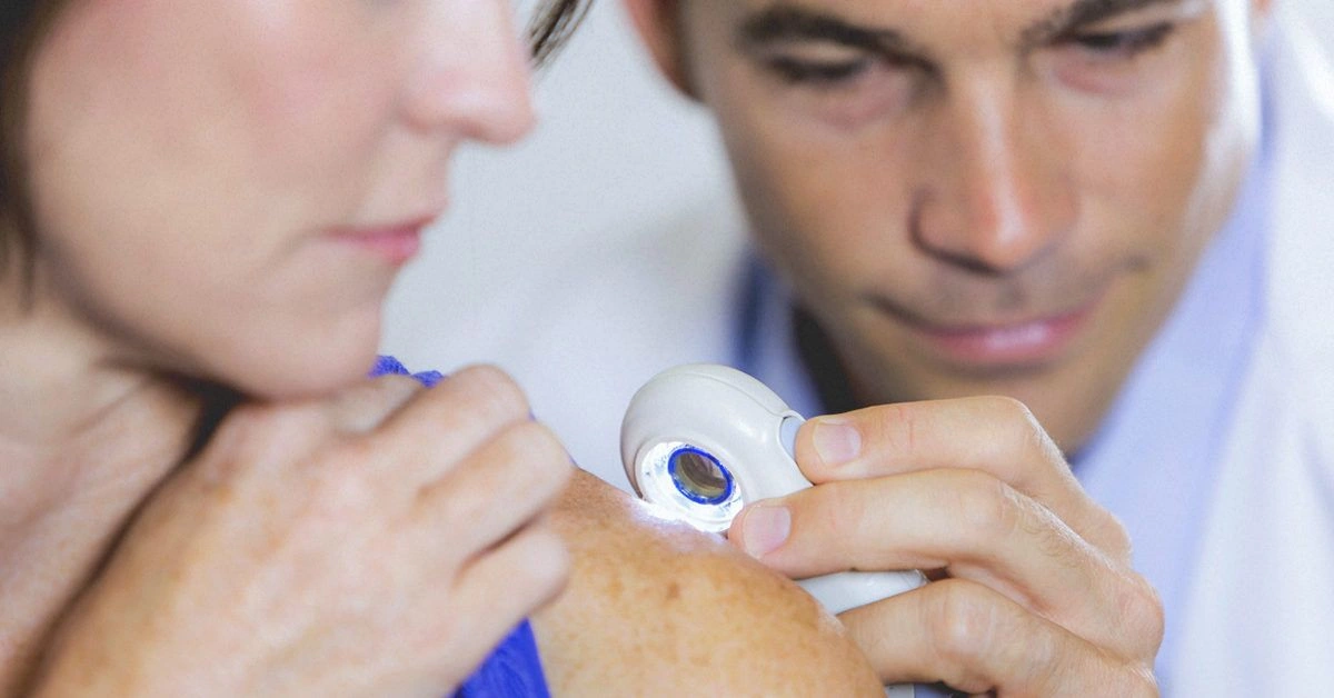
What is a skin neoplasm?
A skin neoplasm is an abnormal growth that appears on the skin. Although the term neoplasm is often used interchangeably with cancer, it also describes noncancerous growths. You may also hear these growths called tumors.
Normally, skin cells divide and grow as needed, and then die when they’re no longer required. Occasionally, however, cells keep proliferating when they shouldn’t, which leads to the formation of neoplasms.
Benign vs. malignant neoplasms
Neoplasms can be categorized in various ways. One primary distinction is whether a neoplasm is benign or malignant.
Benign neoplasms
A benign neoplasm stays localized where it began and does not spread into nearby tissues or distant parts of the body. Still, it can compress or damage surrounding organs and tissues. Benign neoplasms are noncancerous.
Keep in mind that some benign neoplasms may eventually become cancerous. Even with a benign diagnosis, you should monitor the area and report any changes in appearance to your physician promptly.
Examples of benign skin neoplasms include:
- skin tags
- cherry angioma
- dermatofibroma
- epidermoid cyst
- keratoacanthoma
- lipoma
- pyogenic granuloma
- sebaceous hyperplasia
- seborrheic keratosis
Malignant neoplasms
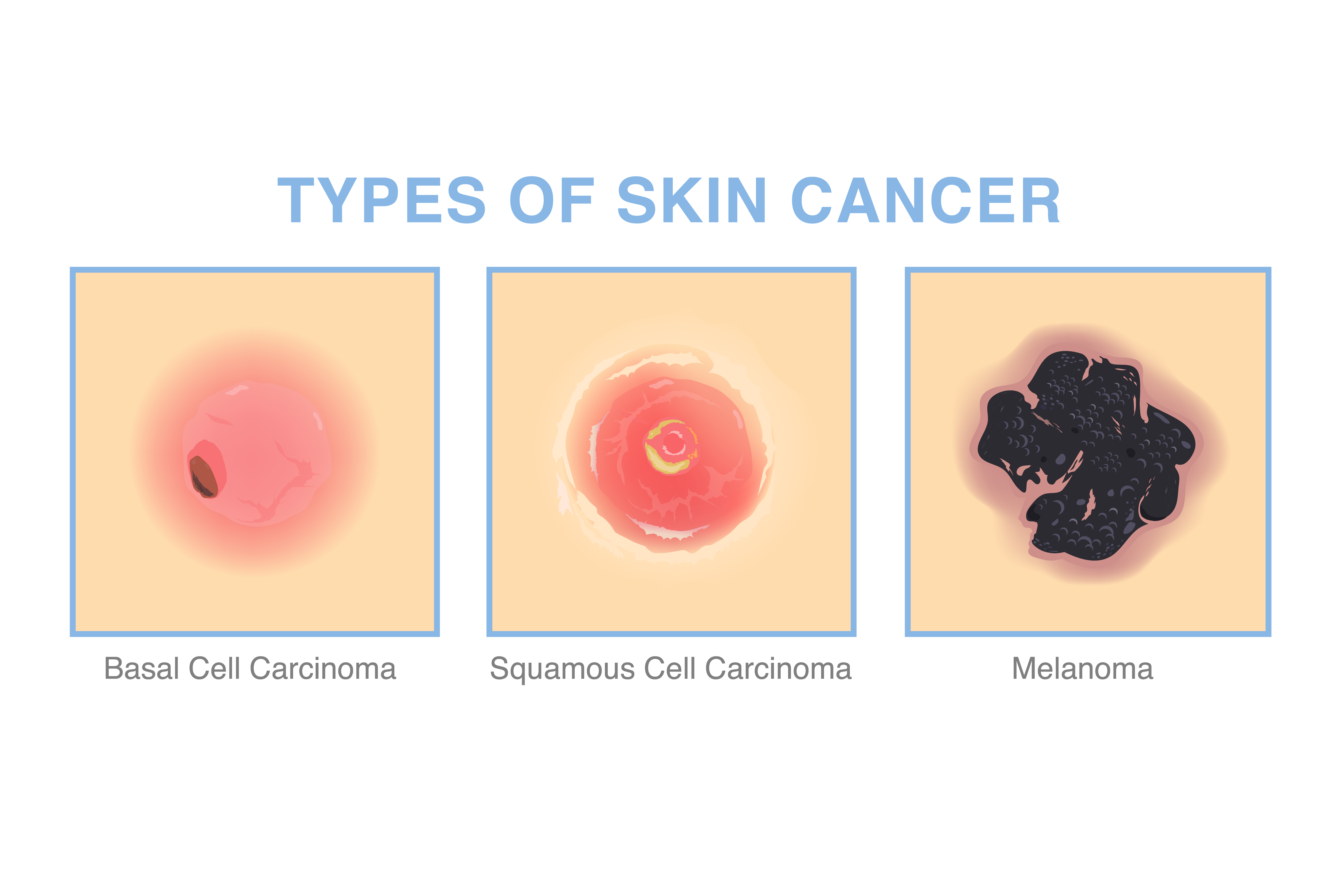
A malignant neoplasm is cancerous. Unlike benign growths, malignant neoplasms grow unchecked and can infiltrate nearby tissues. They may also spread through the body and form secondary tumors.
Common malignant skin neoplasms include:
- basal cell carcinoma
- squamous cell carcinoma
- melanoma
- Merkel cell carcinoma
- Kaposi sarcoma
- lymphoma of the skin
What about precancerous neoplasms?
Some neoplasms are classified as precancerous. While definitions may vary slightly among clinicians, this generally means the growth has the potential to become cancer if not managed. In some cases these lesions resolve on their own, but others may slowly progress to cancer.
For instance, actinic keratosis (AK), also known as solar keratosis, presents as rough, scaly patches on skin that’s been exposed to ultraviolet radiation. AKs can look like warts and may appear pink or flesh-toned. Without treatment, they can evolve into a form of squamous cell skin cancer.
Another frequent precancerous lesion is squamous cell carcinoma in situ, sometimes called Bowen’s disease. The term “in situ” means “in its original place,” indicating the atypical cells are confined to the epidermis, the outermost skin layer. Squamous cell carcinoma in situ often appears as red, scaly patches that may itch. If untreated, it can progress to invasive squamous cell carcinoma.
What is uncertain behavior?
Besides being labeled malignant or benign, some neoplasms are described as having uncertain behavior. This indicates that more information is needed to decide whether the lesion is cancerous or not — in other words, it’s unclear if it can spread.
When a neoplasm is of uncertain behavior, your doctor will probably recommend a biopsy. That involves removing a small piece of tissue from the area to test for cancer.
Check yourself
Whether you currently have a skin neoplasm or not, it’s important to examine your skin regularly for any new lesions. Once a month, stand before a mirror and inspect your entire body, including hard-to-see places like the back of your neck and the soles of your feet.
The American Academy of Dermatology offers a downloadable body map and chart you can use to document any findings. Keeping routine notes helps you track changes over time.
You can use the ABCDE method to help spot melanoma, the most dangerous form of skin cancer. Each letter represents a feature to check:
- Asymmetry. One half of the mole does not match the other.
- Borders. The edges are irregular or notched.
- Color. The lesion shows multiple colors or uneven shading.
- Diameter. The spot is larger than ¼ inch (about the size of a pencil eraser).
- Evolving. The lesion has changed in size, shape, or color.
Other warning signs include:
- a change in the lesion’s texture
- a spot that itches, crusts, scabs, or bleeds
- an open sore that does not heal within three weeks
If you notice any of these features anywhere on your body, see your doctor right away. These signs don’t automatically mean the lesion is cancerous, but prompt evaluation is important. Early detection generally makes treatment easier. Learn more about how to screen yourself for skin cancer.
What should I do if I find a new growth?
If you discover a new growth on your skin, schedule an appointment with a dermatologist. Your primary care provider can refer you if needed.
At the visit, the dermatologist will likely ask about your medical history and habits. Tell them about anything that could raise your risk for skin cancer, such as:
- having lighter skin
- a family history of skin cancer
- past use of tanning beds
- a large number of moles
- skin that burns easily or freckles
- history of sunburns, particularly during childhood
Next, the doctor will examine the lesion. Based on what they see, they may perform a biopsy to determine whether it’s benign or malignant.
The three main types of skin biopsy are:
- Shave biopsy. A small blade is used to shave off the upper layers of skin.
- Punch biopsy. A circular instrument removes a small, round sample from the top and deeper skin layers.
- Excisional biopsy. A scalpel is used to cut out the entire lesion and a margin of surrounding skin.
Your dermatologist will send the tissue sample to a lab for analysis. Once results are available, they’ll review them with you and discuss treatment options if necessary. Learn more about what to expect during a skin cancer biopsy.
The bottom line
A skin neoplasm is an abnormal skin growth. These growths are commonly classified as benign, malignant, or precancerous. Doctors may also describe a lesion as having uncertain behavior if they cannot immediately determine whether it is cancerous.
Regardless of whether you have a diagnosed neoplasm, it’s wise to perform regular skin self-exams to monitor for new growths or changes in existing ones.















Leave a Reply
You must be logged in to post a comment.