Internal organs located on your left side include the transverse and descending colon, the pancreas, as well as the left lung and kidney, among others.
Although the outside of the human body appears mostly symmetrical, the organs housed on the left and right sides differ in both placement and function.
Below is a concise anatomical overview of the left side of your body, which contains the following organs:
- the left hemisphere of the brain
- left eye and ear
- lung
- heart
- adrenal gland
- spleen
- kidney
- stomach
- pancreas
- liver
- transverse colon and descending colon
- reproductive organs
The brain’s left hemisphere
Weighing approximately 3 pounds, the brain is one of the most intricate organs in the human body. While its outer structure looks symmetrical, its functional roles are not evenly divided. It is separated into two hemispheres: left and right.
What it does
The left hemisphere is primarily responsible for language production and word formation, whereas the right hemisphere plays a greater role in abstract reasoning and spatial processing.
That said, research using magnetic resonance imaging (MRI) has shown that the concept of being strictly “left-brained” or “right-brained” is oversimplified. Both hemispheres collaborate and carry out essential neurological functions.
Brain and body
Most nerve pathways between the brain and the body cross to the opposite side. This means the left hemisphere largely governs movements and sensations on the right side of the body. If damage occurs to one side of the brain, such as during a stroke, the opposite side of the body is typically affected.
Left ear
The ears are composed mainly of cartilage and have a curved, shell-like structure.
Each ear is divided into three sections:
- outer ear and ear canal
- middle ear
- inner ear
What it does
The ear detects sound vibrations traveling through the air and distinguishes both pitch (the frequency of sound waves) and volume (the intensity or loudness of sound).
Sound perception
The cochlea, located within the inner ear, houses the organ of Corti. This specialized structure contains sensory hair cells that convert mechanical movement into electrical impulses, which are then transmitted to the brain for interpretation.
Left eye
The eyes measure roughly 1 inch — or 2.5 centimeters (cm) — in diameter.
The main anatomical components of the eye include:
- retina
- cornea
- iris
- ciliary body
- lens
- sclera
What it does
The eyes receive light from the surrounding environment and relay visual information to the brain via the optic nerve, also known as the second cranial nerve.
Various parts of the eye collaborate to properly focus incoming light onto the retina.
The retina contains rods and cones that enable vision in different lighting conditions. Rods are particularly important for low-light vision, while cones help detect color and fine detail.
Cones and rods
The human eye contains approximately 6 million cone cells and 90 million rod cells.
Left lung
Your left lung consists of two lobes, whereas the right lung contains three. This structural difference accommodates the position of the heart on the left side.
What it does
The lungs serve as the primary organs of respiration. They draw in oxygen and expel carbon dioxide. Located within the rib cage, they are composed of soft, spongy tissue that expands and contracts with each breath.
Key structures involved in airflow include:
- bronchi
- bronchiole tubes
- alveoli
The lungs have relatively few pain receptors. As a result, lung-related problems often present with symptoms such as coughing, wheezing, or shortness of breath rather than sharp pain.
Self-cleaning lungs
The lungs contain a self-cleaning, brushlike apparatus made up of cilia that help remove mucus, debris, and potentially harmful particles from the airways.
Heart
The heart is positioned in the center of your chest, slightly to the left. This muscular organ forms the core of your circulatory system and is divided into left and right chambers.
In adults, the heart is roughly the size of a clenched fist: about 5 inches (12 cm) long, 3.5 inches (8–9 cm) wide, and 2.5 inches (6 cm) deep, according to Henry Gray’s 1918 “Anatomy of the Human Body.”
What it does
The heart propels blood through a network of blood vessels. Oxygen-rich blood is delivered to the brain and body tissues, then returns to the lungs to receive more oxygen.
The heart has four chambers:
- two upper chambers called atria, right and left. The right atrium collects oxygen-depleted blood returning from the body (except the lungs). The left atrium receives oxygenated blood from the lungs.
- two lower chambers called ventricles, right and left. The right ventricle pumps oxygen-depleted blood to the lungs. The left ventricle sends oxygen-rich blood to the rest of the body (excluding the lungs).
The circulatory system also includes:
- arteries, which carry oxygen-rich blood away from the heart
- capillaries, which enable exchange of nutrients, gases, and waste
- veins, which return oxygen-depleted blood to the heart
Reading your heart
Blood pressure reflects how effectively the heart pumps blood.
The top number represents arterial pressure when the heart contracts and pushes blood out of the ventricles.
The bottom number reflects pressure in the arteries when the heart relaxes between beats.
A normal blood pressure reading is typically 120 or lower for the top number and 80 or lower for the bottom number.
Adrenal gland
You have two adrenal glands, each sitting on top of a kidney.
What it does
Despite their small, triangular shape, the adrenal glands are crucial for immune function, metabolism, and stress regulation.
The pituitary gland in the brain directs adrenal activity by releasing hormones that regulate the endocrine system.
The adrenal gland consists of two main regions:
- The adrenal cortex, the outer layer, produces aldosterone and cortisol.
- The adrenal medulla, the inner portion, releases epinephrine (adrenaline) and norepinephrine (noradrenaline), which control the fight-or-flight response.
Subtle signs from hormones
If hormone production becomes excessive or insufficient, symptoms may be mild at first. Low blood pressure, dizziness, or unusual fatigue can signal an imbalance. Persistent symptoms should be evaluated by a healthcare professional.
Spleen
The spleen rests beneath the diaphragm and behind the upper left ribs. About 5 inches (13 cm) long or smaller and purple in color, it is protected by the rib cage.
What it does
As a component of the lymphatic system, the spleen filters blood, recycles red blood cells, and releases white blood cells known as lymphocytes to support immune defense.
It also produces substances that help reduce inflammation and assist with healing.
The replaceable spleen
It is possible to live without a spleen. If removal is necessary due to injury or disease, the liver and lymph nodes can compensate for many of its key roles.
Left kidney
You have two kidneys situated below your rib cage on either side of the spine. The left kidney is usually slightly larger than the right.
What it does
The kidneys remove waste and surplus fluid from the bloodstream, producing urine. They also maintain proper electrolyte balance and contribute to blood pressure regulation and red blood cell production.
Each kidney contains around 1 million nephrons and filters approximately 200 liters of fluid daily.
A nephron consists of a renal corpuscle (including the glomerulus) and a tubule. The glomerulus filters blood, while the tubule reabsorbs essential substances and eliminates waste.
One kidney alone can adequately sustain normal body function if healthy.
Kidneys in history
Ancient Egyptians documented knowledge of the kidneys in a papyrus dating from 1500 B.C. to 1300 B.C.
Stomach
The stomach is positioned in the upper, middle-left region of the abdomen, beneath the diaphragm and in front of the spleen.
What it does
The stomach temporarily stores food and liquids and initiates digestion through the action of gastric acids and enzymes.
After approximately 2 to 5 hours, partially digested contents pass into the small intestine for further breakdown and absorption.
Expandable folds called rugae allow the stomach to stretch and accommodate varying amounts of food.
Protective mucus
Because stomach acid has a pH between 1 and 2, a protective mucus lining is essential to shield the stomach wall from corrosion.
Pancreas
The pancreas lies deep within the abdomen, behind and slightly below the stomach.
What it does
This gland produces digestive enzymes that help break down fats, starches, and proteins in the small intestine.
It also secretes insulin and glucagon, hormones responsible for maintaining balanced blood sugar levels.
Hidden symptoms
More than 37,000 new cases of pancreatic cancer are diagnosed annually in the United States, according to the National Pancreas Foundation. Yellowing of the skin without additional symptoms may be an early sign.
Left lobe of the liver
The majority of the liver is located on the right side, but a smaller left lobe extends across the midline above the stomach.
The liver weighs about 3 pounds on average, according to the CDC.
What it does
The liver plays a central role in metabolism, energy production, detoxification, and nutrient processing.
It regulates blood chemistry, stores vitamins and minerals, and produces bile to aid fat digestion. It also breaks down carbohydrates, fats, and proteins.
Bile is excreted in feces, while blood waste products are filtered by the kidneys and eliminated in urine.
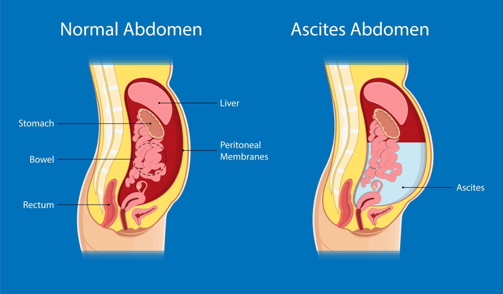
The liver has a remarkable ability to regenerate its cells, but it is essential for survival. For visual references of abdominal swelling linked to liver conditions, see Pictures of swollen abdomen due to liver disease and Cirrhosis liver belly images female.
Made of lobes
Anatomically, the liver has four lobes. Under the Couinaud classification system, it is divided into eight functional segments, each with its own bile duct.
Transverse and descending colon
The colon, or large intestine, forms an inverted U shape over the small intestine.
The ascending colon is on the right, the transverse colon spans the top, and the descending colon runs down the left side.
What it does
The descending colon stores digestive waste and gradually converts liquid stool into a more solid form before elimination.
It connects to the sigmoid colon, which is named for its S-shaped curve.
The end of the line
The descending colon measures about 3.9 to 5.9 inches (10 to 15 cm) in length and roughly 2.5 inches (6.3 cm) in width. The entire colon extends about 5 feet (1.5 m), according to the National Cancer Institute.
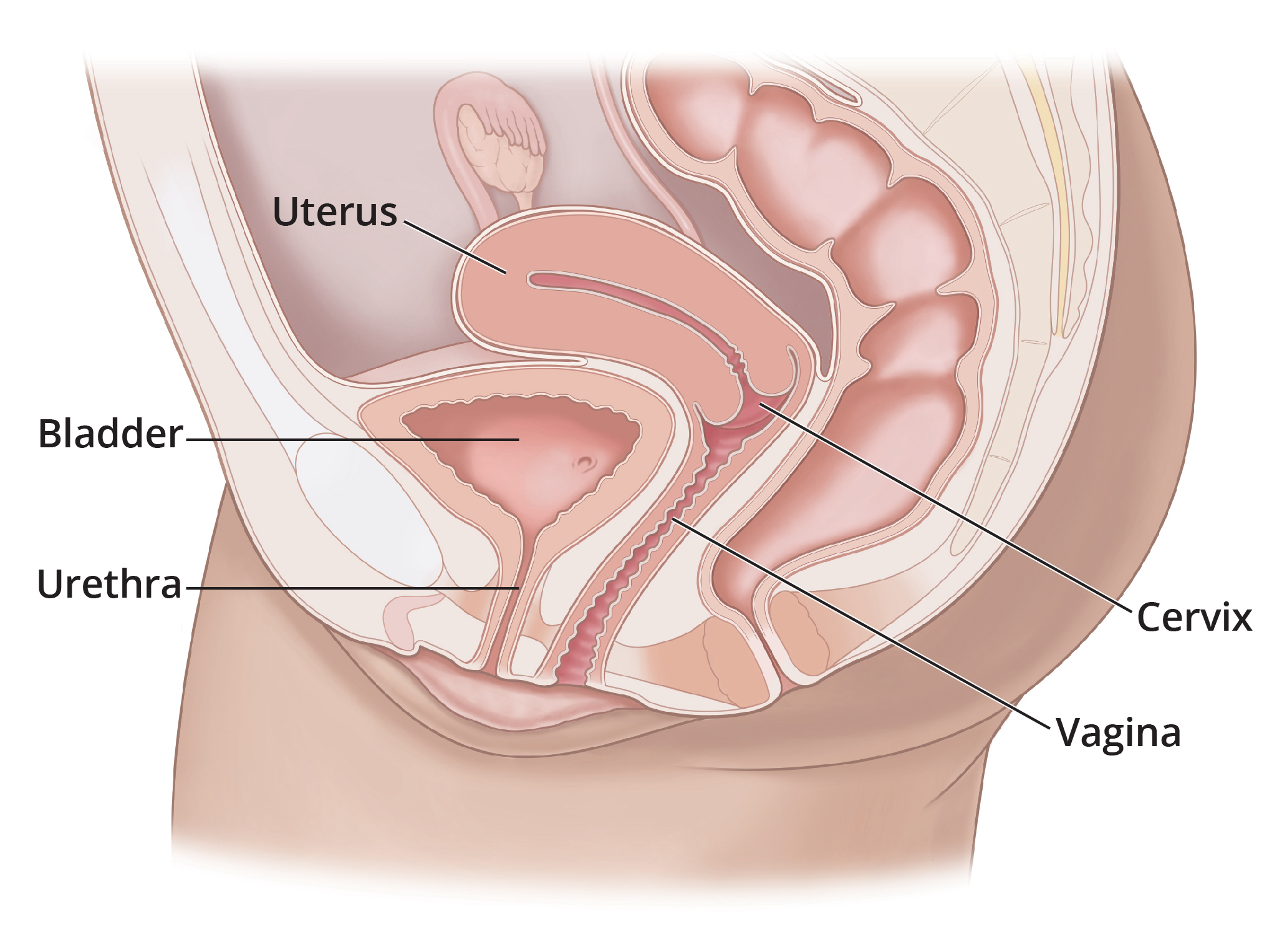
Female and male reproductive organs on the left
Left ovary
Each ovary, approximately almond-sized, is located on either side of the uterus.
What it does
During the reproductive years, ovulation typically occurs once per month, releasing an egg around the midpoint of a 28-day menstrual cycle.
The ovaries also secrete estrogen and progesterone, hormones vital for reproductive health.
Left fallopian tube
The fallopian tube connects the ovary to the uterus and provides the pathway for the egg.
What it does
Fertilization usually occurs within the fallopian tube when sperm meets the egg.
Left testis
The testes are located in the scrotum and are oval-shaped, measuring about 1.8 to 2 inches (3 to 5 cm) long.
What it does
The testes produce sperm and the androgen hormone testosterone. Sperm travel through a thin tube to the urethra for ejaculation.
The takeaway
Your body is an intricate biological system with numerous specialized organs. Many of these critical structures are positioned on the left side.
Situs inversus: left and right reversal
Approximately 1 in 10,000 individuals are born with their internal organs reversed, a condition known as complete situs inversus. This phenomenon was first described in 1788 by Matthew Baillie, MD.


Leave a Reply
You must be logged in to post a comment.