

Overview
Dacryocystitis is an infection of the tear sac (lacrimal sac) located at the inner corner of the eye. This sac is part of the pathway through which tears exit the eye.
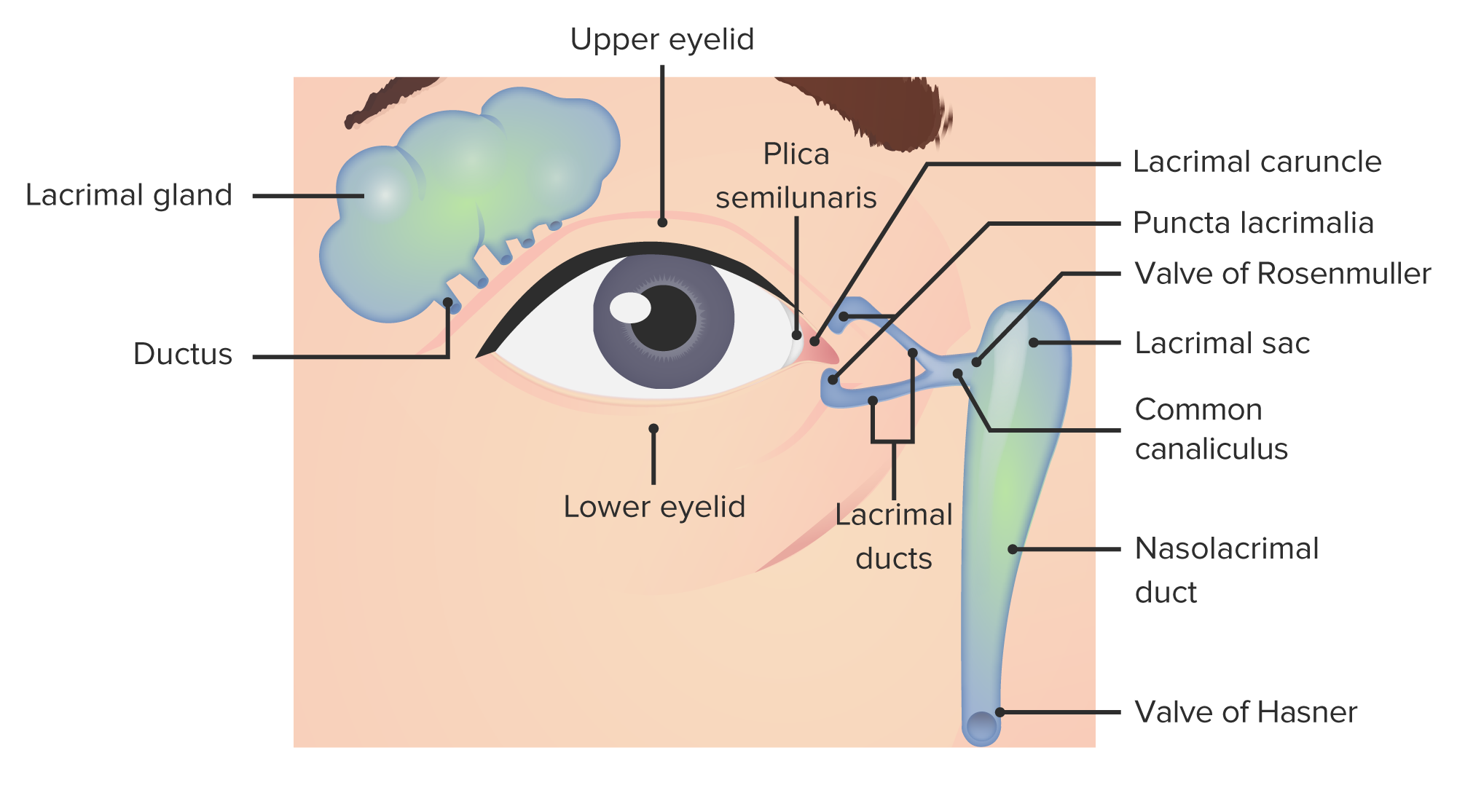
Tears keep the eyes moist, clear, and help prevent bacterial growth. The lacrimal glands beneath the upper eyelids produce tears that move across the eye surface via tiny openings. Each blink spreads the tears over the eye. Excess fluid drains out through small openings called puncta at the corners of the upper and lower eyelids. From there, tears pass into the nasolacrimal sac, travel down the tear duct (lacrimal duct), and empty into the back of the nose.
Infections most commonly occur when the lacrimal duct becomes blocked, allowing bacteria to accumulate in the tear sac.
A rapidly starting infection is termed acute dacryocystitis. When the infection persists over time, it is referred to as chronic dacryocystitis.
Symptoms
Signs of acute dacryocystitis include:
- pain, redness, and swelling at the inner corner of the eye
- excessive tearing
- localized swelling next to the nose
- redness of the eye
- discharge of pus or mucus from the corner of the eye
- fever
Chronic dacryocystitis often produces milder signs. You may have persistent tearing and some discharge, with little or no noticeable swelling.
Your healthcare provider will make a diagnosis based on your symptoms and an eye exam. Occasionally imaging, such as CT scans, is used to identify the cause of the blockage.
Treatment
The primary treatment for dacryocystitis is antibiotics, which eradicate the bacteria responsible for the infection. Antibiotics are usually taken orally, but severe cases may require intravenous administration. Your clinician may also recommend antibiotic eye drops or ointment.
Applying warm compresses to the affected eye several times a day can help ease pain and reduce swelling.
Once the infection has resolved, a surgical procedure called dacryocystorhinostomy (DCR) is sometimes needed. This operation creates a new passage around the obstructed duct, often by removing a small area of bone, allowing tears to drain directly from the lacrimal sac into the nose. Enlarging the drainage route helps prevent recurrent infections. The procedure can often be performed endoscopically through very small incisions.
Causes
The infection typically begins due to a blockage of the tear duct. Possible contributors to this obstruction include:
- trauma to the nose or eye, such as a fractured nose
- nasal polyps, which are growths inside the nose
- sinus inflammation or infection
- prior nasal or sinus surgery
- a foreign body lodged in the duct
- malignant growths
Dacryocystitis is particularly common in infants, who may be born with a blocked tear duct; this is known as congenital dacryocystitis.
Middle-aged women are more susceptible than men, since their tear ducts tend to be naturally narrower. The condition also becomes more frequent with advancing age.
Other factors that increase the risk of dacryocystitis include:
- a deviated septum, where the thin wall between the nostrils is displaced, making one nasal passage smaller
- rhinitis, or inflammation of the nasal mucous membrane
- inferior turbinate hypertrophy, which is swelling of one of the bony structures inside the nose that filter and humidify inhaled air
Prevention
You can reduce the risk of future infections by undergoing dacryocystorhinostomy to enlarge the obstructed duct.
If you or your child frequently experience tear duct infections, one preventive measure is to manually drain the tear sac. After washing your hands, apply a warm, damp washcloth over the tear sac. Gently place a finger at the corner of the eye near the nose and press on the sac. This may release fluid or pus. Follow by reapplying the warm compress.
Complications
An untreated acute infection can progress to a chronic condition. In infants with congenital dacryocystitis, the infection can extend into the eye socket, potentially causing severe, life-threatening complications such as:
- brain abscess, a localized collection of pus within the brain
- meningitis, inflammation of the protective membranes covering the brain and spinal cord
- sepsis, a systemic inflammatory response to infection

Outlook
Acute dacryocystitis generally responds quickly to antibiotic therapy. Many infants with the congenital form outgrow the blockage by around age 1 as the tear duct naturally widens.
Chronic lacrimal sac infections can be more difficult to manage and may require surgical intervention to widen the tear drainage pathway.





















Leave a Reply
You must be logged in to post a comment.