Imagine you’re at a beach party, the sun is shining, and a few weeks later a pink‑purple, ring‑shaped rash pops up on the back of your neck. You brush it off as a sunburn, but it reappears after every sunny outing, never really healing and sometimes even spreading. That stubborn, non‑scarring rash could be subacute cutaneous lupus (SCLE) – a skin‑focused form of lupus that often hides in plain sight.
In the next few minutes we’ll walk through what SCLE really is, why it shows up, how doctors pin it down, and what you can do to keep it under control. Grab a cup of tea, settle in, and let’s demystify this lupus skin disease together.
What Is SCLE?
One‑sentence definition
SCLE is a photosensitive, non‑scarring rash variant of cutaneous lupus that typically appears on sun‑exposed areas of the body.
How SCLE differs from other lupus skin diseases
Picture lupus as a family of skin conditions. Discoid lupus (DLE) gives you thick, scarring plaques, while systemic lupus erythematosus (SLE) can involve anything from kidneys to the heart. SCLE sits in the middle: it’s skin‑only for most people, but a noticeable chunk (about 15‑30 %) may later develop systemic involvement.
Who gets SCLE?
The classic demographic is a white woman between 15 and 40 years old, representing roughly 10 % of all lupus cases. Genetics play a subtle role, but most often the trigger is something external – especially sunlight.
Quick‑reference comparison
| Feature | SCLE | Discoid Lupus (DLE) | Systemic Lupus (SLE) |
|---|---|---|---|
| Rash type | Annular, poly‑cyclic, non‑scarring | Disc‑shaped, scarring | Variable, may include rash |
| Photosensitivity | ≈ 80 % of patients | ≈ 50 % | ≈ 70 % |
| Risk of systemic disease | 15‑30 % progress to SLE | Rare | Universal |
| Typical sites | V‑neck, forearms, upper back | Face, scalp, ears | Any organ |
SCLE Symptoms Overview
Classic skin patterns
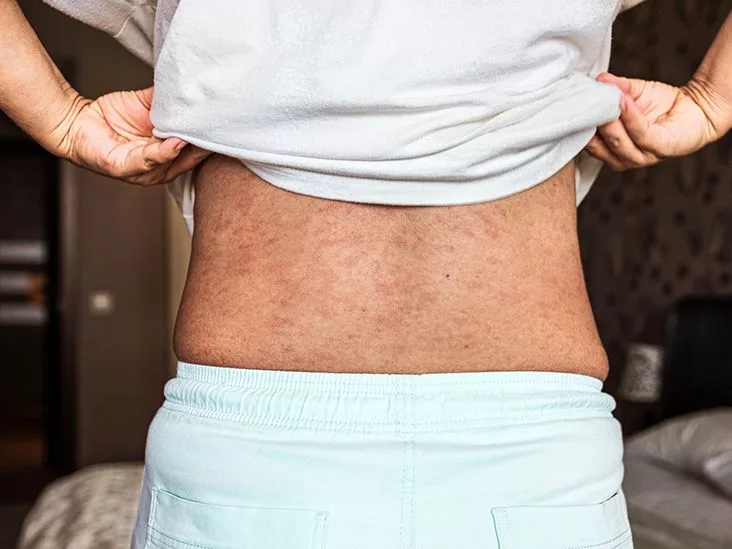
The hallmark is an annular or poly‑cyclic plaque with a raised, reddened border and a lighter centre – essentially a “ring‑rash” that may be slightly scaly. These lesions love the sun, so you’ll see them on the “V” of the neck, the forearms, or the upper back.
Accompanying signs
Most people feel fine, but a few notice fatigue, mild joint aches, or flu‑like malaise. Those systemic clues are usually subtle, which is why many patients assume the problem is only skin‑deep.
When to suspect SCLE vs. other rashes
- Rash appears after sun exposure and improves with shade.
- Lesions are non‑scarring and tend to recur in the same locations.
- Blood tests reveal positive anti‑Ro/SSA or anti‑La/SSB antibodies (more on that later).
A real‑world story
Sarah, a 27‑year‑old graphic designer, first noticed a pink ring on her right forearm after a weekend hike. It faded in a week, only to reappear a month later on her neckline. After a visit to her dermatologist, blood work showed anti‑Ro antibodies, confirming SCLE. Today she manages the condition with sunscreen, hydroxychloroquine, and a few lifestyle tweaks.
Why Does SCLE Occur?
Autoimmune background
In SCLE the immune system mistakenly attacks proteins in the skin. The most common serological markers are anti‑Ro/SSA and anti‑La/SSB antibodies – you’ll often see these mentioned in lab reports.
Environmental triggers
Ultraviolet (UV) light is the biggest culprit; about 80 % of patients report a clear link between sunlight and flare‑ups. Certain medications – thiazide diuretics, calcium‑channel blockers, and some antifungals – can also provoke SCLE. If you’ve started a new drug and notice a rash, it’s worth discussing with your doctor.
Genetic and familial links
Family history of lupus or other autoimmune diseases modestly increases risk, but most cases arise without a known genetic predisposition.
Systemic lupus (SLE) risk
While many live with skin‑only disease, roughly one‑third may later develop systemic involvement. Warning signs include persistent joint pain, unexplained fever, or new organ‑specific symptoms (e.g., kidney issues). Ongoing monitoring is key.
Clinician’s checklist (you can use it at home)
- Recent sun‑intense activity?
- Started a new medication?
- Positive anti‑Ro/SSA or anti‑La/SSB?
- Family history of autoimmunity?
How Is SCLE Diagnosed?
Clinical examination
Dermatologists look for the classic distribution (V‑neck, forearms, upper back) and the characteristic annular, non‑scarring plaques. They also ask about photosensitivity and medication history.
Laboratory work‑up
Typical labs include:
- ANA (antinuclear antibody) – often positive.
- ENA panel – specifically anti‑Ro/SSA and anti‑La/SSB.
- Basic metabolic panel and CBC to screen for systemic involvement.
Skin biopsy
If the visual clues aren’t decisive, a 4‑mm punch biopsy can confirm SCLE. Histology usually shows interface dermatitis with basal keratinocyte vacuolization – a pattern you’ll read about on DermNet NZ.
Differential diagnosis
Other conditions that can mimic SCLE include psoriasis, drug eruptions, dermatomyositis, and even allergic contact dermatitis. The biopsy and antibody profile help separate them.
Diagnostic flowchart (simplified)
- History + photo review →
- Physical exam →
- Blood work (ANA, anti‑Ro/SSA, anti‑La/SSB) →
- If needed, skin biopsy →
- Rule out other causes → Diagnosis confirmed.
Effective SCLE Treatment
First‑line: Sun protection
Think of sunscreen as the most powerful prescription you’ll ever fill. Choose a broad‑spectrum SPF 30 or higher, reapply every two hours, and wear UV‑blocking clothing, wide‑brim hats, and sunglasses. Even on cloudy days, UV rays can trigger a flare.
Topical therapy
For active lesions, low‑ to mid‑potency corticosteroids (hydrocortisone 1 %‑2.5 % or triamcinolone) are the go‑to. If you need something gentler for the face or neck, calcineurin inhibitors like tacrolimus or pimecrolimus work well and avoid skin thinning.
Systemic agents
When the rash is widespread or persistent, oral medication steps in. The gold standard is hydroxychloroquine (200‑400 mg daily). It’s backed by decades of research and is usually well tolerated, though you’ll need an annual eye exam to monitor retinal health.
If hydroxychloroquine isn’t enough, rheumatologists may add methotrexate, azathioprine, or mycophenolate mofetil – each with its own risk profile that your doctor will explain.
Drug‑induced SCLE
If a medication is the trigger, the simplest cure is stopping the offending drug (under your physician’s guidance, of course). Many patients see rapid improvement after the culprit is removed.
Monitoring for systemic progression
Because of the systemic lupus risk, schedule regular check‑ups: repeat blood work every 6‑12 months, and report any new joint pain, fatigue, or organ‑specific symptoms right away.
Topical vs. systemic: quick look
| Therapy | When to use | Typical dose | Onset of relief | Common side‑effects |
|---|---|---|---|---|
| Topical steroid | Isolated flare | 0.05‑0.1 % cream | 1‑2 weeks | Skin thinning, telangiectasia |
| Calcineurin inhibitor | Face/neck, steroid‑sparing | 0.1 % ointment | 2‑4 weeks | Burning, itching |
| Hydroxychloroquine | Persistent/widespread disease | 200‑400 mg/day | 4‑12 weeks | Retinal toxicity (annual eye exam) |
| Methotrexate | Refractory SCLE | 7.5‑25 mg weekly | 6‑12 weeks | Hepatotoxicity, cytopenia |
Living With SCLE
Daily sun‑safety routine
Make sunscreen part of your morning ritual: apply 15 minutes before heading out, re‑apply after swimming or sweating, and don’t forget often‑missed spots like the ears and back of the hands. There are handy smartphone apps that give you the real‑time UV index – a fun way to stay vigilant.
Skin‑care basics
Use gentle, fragrance‑free cleansers, and follow up with a rich, non‑comedogenic moisturizer. A well‑hydrated skin barrier tolerates UV exposure better and heals faster.
Medication adherence
Hydroxychloroquine works best when taken consistently. Set a daily alarm, pair the pill with another routine (like brushing your teeth), and keep a medication diary if that helps.
When to call your doctor
- New or worsening rash despite sunscreen.
- Joint pain, unexplained fever, or fatigue that lasts more than two weeks.
- Any signs of organ involvement – swelling, shortness of breath, or changes in urine.
Support and community
Living with a chronic skin condition can feel isolating. Online forums, local lupus support groups, and patient‑education pages from the American College of Rheumatology provide a space to share tips, ask questions, and find encouragement.
Key Takeaways
Subacute cutaneous lupus is a photosensitive, non‑scarring rash variant of lupus that primarily targets sun‑exposed skin but carries a modest risk of becoming systemic. Recognizing the classic annular lesions, ordering the right labs (especially anti‑Ro/SSA and anti‑La/SSB), and confirming with a skin biopsy when needed are the cornerstones of diagnosis. Effective management blends diligent sun protection, topical anti‑inflammatories, and, for many, the oral workhorse hydroxychloroquine. Lifestyle tweaks—sunscreen, gentle skin‑care, and regular medical follow‑up—can keep the disease in check and preserve quality of life.
Remember, you’re not alone on this journey. If you’ve experienced a tricky rash, noticed a pattern, or just have questions about SCLE, share your story in the comments below. Let’s learn from each other and turn uncertainty into confidence.


Leave a Reply
You must be logged in to post a comment.