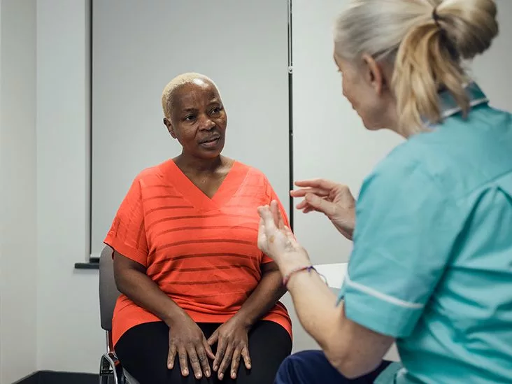
Seeing a drop of blood after you’ve been blissfully period‑free can feel like a tiny alarm clock going off at the worst possible moment. The truth is that postmenopausal bleeding can be a sign of cancer, but in more than nine‑tenths of cases it comes from something completely harmless. Knowing the difference, understanding the next steps, and taking action early is the best way to keep calm and stay healthy.
In the next few minutes I’ll walk you through the red‑flag signs, the most common benign reasons, what doctors actually do when you call, and the lifestyle tweaks that can lower your risk. Think of this as a friendly coffee chat—no jargon, just clear, caring information you can actually use.
Spotting Cancer Risks
Is any post‑menopausal bleeding a red flag?
Short answer: yes. Even a single spotting episode should trigger a call to your GP. According to a Harvard Health study, more than 90 % of women who were later diagnosed with endometrial (uterine) cancer first noticed some form of bleeding.
What amount or pattern should worry me?
It isn’t the volume that matters most—it’s the fact that bleeding is occurring at all after a year of menopause. Light spotting, brown discharge, or a few drops on your underwear are all worth checking. The JAMA Internal Medicine analysis (2019) found that the sheer presence of bleeding, regardless of heaviness, was the strongest predictor of cancer.
Can a single spot be ignored?
While a one‑off spot is often caused by atrophic vaginitis (thin, dry vaginal tissue), it’s still safest to have a clinician look. Think of it like a car’s “check engine” light—sometimes it’s a tiny sensor, other times it warns of a bigger problem.
When should I call the doctor right away?
Call ASAP if you notice any of these:
- Bleeding that won’t stop after a few days
- Heavy flow, clots, or bright red streams
- Accompanying pain, pelvic pressure, or unexplained weight loss
- Fever, foul odor, or discharge that looks like pus
These “red‑flag” symptoms raise the suspicion of cancer and speed up the diagnostic process.
Quick‑Reference Risk Table
| Bleeding pattern | Typical benign cause | Cancer risk* |
|---|---|---|
| One‑off spotting | Atrophic vaginitis | Low |
| Persistent spotting > 2 weeks | Polyps / atrophy | Moderate |
| Heavy bleeding or clots | Fibroids, polyps | Higher |
| Bleeding + pain/weight loss | Possible malignancy | High |
*Risk percentages are summarised from National Cancer Institute (NCI) and Harvard data (2023‑2025).
Common Benign Causes
Atrophic vaginitis & endometrial atrophy
After menopause, estrogen levels drop, and the lining of the vagina and uterus can become very thin. Tiny blood vessels break easily, creating that pink‑brown “spotting” most women experience. It’s uncomfortable, but it’s usually not cancerous.
Uterine polyps and fibroids
Polyps are small, non‑cancerous growths on the inner wall of the uterus or cervix. Fibroids are larger, muscular tumors that can also bleed. Both often show up on a routine ultrasound and can be removed with a simple hysteroscopic procedure.
Hormone‑replacement therapy (HRT) effects
If you’ve started HRT, especially estrogen‑only regimens, a little breakthrough bleeding is fairly common during the first few months. Adding a progestin or adjusting the dose usually settles things. The NHS notes that any bleeding while on HRT still warrants a check‑up, just to be sure.
Medications and other triggers
Anticoagulants (blood thinners), tamoxifen (used for breast cancer), and selective estrogen receptor modulators (SERMs) can all provoke bleeding. If you’re on any of these, let your doctor know—often a dosage tweak prevents the symptom.
Why does estrogen excess cause polyps?
Estrogen stimulates the uterine lining to grow. In women with obesity, excess estrogen is produced by fat tissue, which can lead to overstimulation and formation of polyps or hyperplasia (thickened lining). The National Cancer Institute explains this link in depth.
Doctor’s Diagnostic Pathway
Step 1: Medical history & symptom checklist
Your doctor will ask when the bleeding started, how much, any triggers (sex, trauma), medications, family cancer history, and whether you’re on HRT. This conversation is the foundation for everything that follows.
Step 2: Physical exam & pelvic inspection
A gentle pelvic exam lets the clinician look for visible polyps, cervical lesions, or signs of atrophy. It can feel awkward, but it’s quick—think of it as a routine car service, just a little more personal.
Step 3: Transvaginal ultrasound (TVUS)
The ultrasound measures the thickness of the endometrial lining. An endometrial echo complex of ≤ 4 mm is generally considered low‑risk for cancer (ACOG guideline). Anything thicker usually triggers the next step.
Step 4: Endometrial sampling / biopsy
If the lining is > 4 mm, a small pipelle device is passed through the cervix to suction a tissue sample. The pathologist looks for atypical cells under a microscope. This office‑based biopsy replaces the older, more invasive dilation & curettage (D&C) in most cases.
Step 5: Hysteroscopy (optional)
When polyps or fibroids are suspected, a thin camera is inserted through the cervix to visualise the uterine cavity directly. The doctor can often remove the growth at the same time, avoiding a separate surgery.
Diagnostic Flowchart (text version)
Bleeding → History → TVUS (≤4 mm?) → No cancer → Observe/Treat Benign → >4 mm → Biopsy → Cancer? → Referral → Treatment
Higher Cancer Risk
Age and average diagnosis
Endometrial cancer most often appears around age 60. While menopause typically happens in the early‑50s, the risk climbs steadily after you hit the mid‑60s.
Obesity and estrogen
Carrying an extra 30 kg (about 66 lb) can double or even quadruple your cancer risk. Fat tissue turns androgen into estrogen, feeding the uterine lining constantly. Losing even 5‑10 % of body weight can lower that risk noticeably.
Chronic anovulation & PCOS
If your ovaries don’t release an egg regularly (as in polycystic ovary syndrome), you’re exposed to unopposed estrogen for longer periods—another pathway to hyperplasia and cancer.
Tamoxifen, unopposed estrogen HRT, and genetics
Long‑term tamoxifen use, estrogen‑only hormone therapy, and hereditary conditions like Lynch syndrome dramatically increase the odds. Women with Lynch syndrome have a 10‑40× higher risk, so annual surveillance (often TVUS) is recommended.
Risk‑Factor Comparison Table
| Risk factor | Relative risk (RR) | Typical mitigation |
|---|---|---|
| BMI ≥ 30 kg/m² | 2‑4× | Weight loss, balanced diet, regular activity |
| Unopposed estrogen HRT | 3‑5× | Add progestin or switch therapy |
| PCOS / chronic anovulation | 2‑3× | Metformin, hormonal regulation, weight control |
| Lynch syndrome | 10‑40× | Genetic counseling, annual TVUS/endometrial biopsy |
Prevention & Early Detection
Stay vigilant with regular pelvic care
Even if you feel fine, an annual pelvic exam gives your clinician a chance to spot subtle changes early. Remember the golden rule: any bleeding after menopause deserves a prompt GP visit—ideally within two weeks.
Maintain a healthy weight
Small, sustainable changes work best. Swap sugary drinks for water, add a brisk 30‑minute walk most days, and include a protein‑rich snack to keep cravings at bay. The CDC reports that every 5 % reduction in body weight can cut estrogen‑driven cancer risk by about 15 %.
Review hormone therapy with a specialist
If you’re on HRT, ask your doctor whether a progestin add‑back or a different formulation could minimise bleeding. Many women find that switching from estrogen‑only pills to a combo patch eliminates spotting completely.
Screen high‑risk women
Women with a family history of endometrial or colorectal cancer, especially those with known Lynch syndrome, should discuss annual transvaginal ultrasound and possibly endometrial biopsy with their specialist. Early detection leads to a 95 % five‑year survival rate (NCI).
Quick‑Check List (your personal action plan)
- Notice any vaginal bleeding after a year of no periods? Call your GP.
- Track the pattern: amount, duration, accompanying symptoms.
- Ask about a transvaginal ultrasound; a thin lining (≤ 4 mm) is reassuring.
- If you’re on HRT, discuss dosing or adding progestin.
- Maintain a BMI under 25 kg/m² if possible; every kilogram counts.
- For family‑history or genetic concerns, schedule a genetics referral.
Quick‑Check Summary
Bleeding after menopause is a signal, not a sentence. Most of the time the cause is benign—atrophic vaginitis, polyps, or a temporary HRT side effect. Yet because more than 90 % of women who later develop endometrial cancer firstly notice bleeding, the safest move is to get checked promptly. The diagnostic pathway is straightforward: history, pelvic exam, transvaginal ultrasound, and—if needed—a biopsy. Knowing your personal risk factors (age, weight, hormonal meds, genetics) helps guide how aggressively you and your doctor pursue testing.
By staying aware, keeping a healthy lifestyle, and partnering with a trusted clinician, you dramatically improve the odds that any cancer, should it ever appear, will be caught early—when treatment success rates soar above 95 %. So the next time you see a speck of blood, don’t panic, but don’t ignore it either. Pick up the phone, make that appointment, and take back control of your health.
Have you or someone you know experienced postmenopausal bleeding? How did the journey unfold? Share your story in the comments—your experience could be the reassurance another woman needs today.


Leave a Reply
You must be logged in to post a comment.