A hepatobiliary iminodiacetic acid scan, commonly called a HIDA scan, is an imaging exam used to visualize organs such as the liver, gallbladder, bile ducts, and portions of the small intestine.
A HIDA, or hepatobiliary, scan is a diagnostic imaging procedure. It’s used to obtain pictures of the liver, gallbladder, bile ducts, and small intestine to help identify medical issues affecting those organs. Bile is a fluid that aids in the digestion of fats.
ADVERTISEMENT
Conditions a HIDA scan can help diagnose
HIDA scans assist in diagnosing a range of conditions, including:
inflammation of the gallbladder, known as cholecystitis
obstructions of the bile ducts
congenital bile duct disorders, for example biliary atresia, a rare condition seen in infants
postoperative complications, such as bile leaks and fistulas, which are abnormal connections between organs
Preparing for a HIDA scan
There are some specific steps to prepare for a HIDA scan:
Fast for four hours before the HIDA scan. Your physician may permit clear liquids.
Tell your doctor about all medications and supplements you take.
Inform your physician if you are pregnant or breastfeeding.
When you arrive at the hospital or imaging center, a technologist will ask you to:
change into a hospital gown
remove all jewelry and metal items before the exam
ADVERTISEMENT
What happens during a HIDA scan
Here’s what typically occurs during a HIDA scan:
An imaging technologist will have you lie on a table and remain very still. They will place a scanning camera above your abdomen.
The technologist will insert an IV (intravenous) needle into a vein in your arm or hand.
A radioactive tracer will be injected into the IV so it can enter your bloodstream.
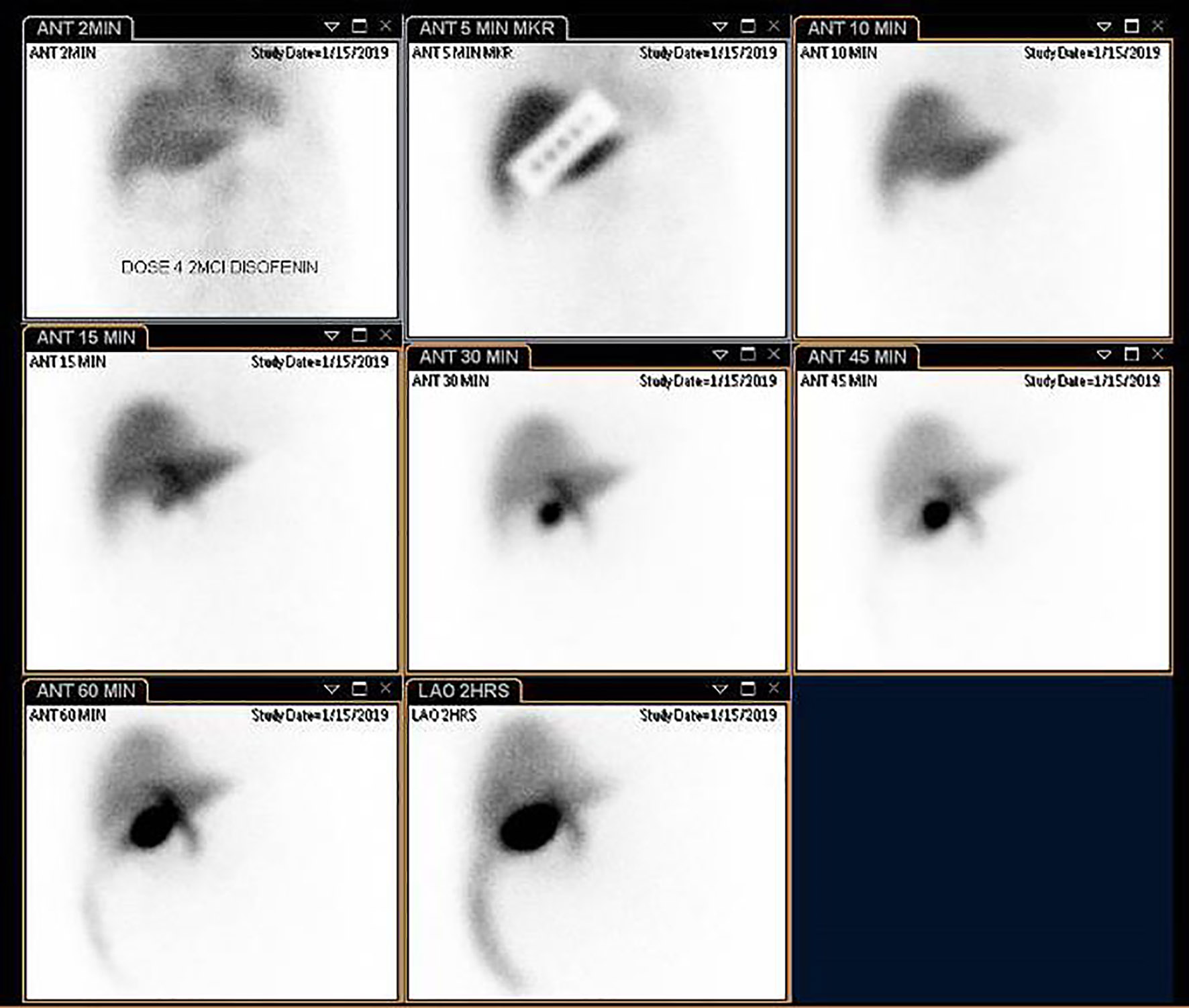
The tracer travels through the bloodstream to your liver, where cells that produce bile take it up. The tracer then moves with bile into the gallbladder, through the bile ducts, and into the small intestine.
The technologist will operate the camera to capture images of the tracer as it moves through these organs.
In some cases, the technologist may administer a pain-relief medication such as morphine via the IV. This can help encourage the tracer to enter the gallbladder.
HIDA scan with CCK
Your provider may request a HIDA scan with CCK (cholecystokinin), a hormone that stimulates the gallbladder to contract and release bile. If ordered, the technologist will give CCK by mouth or through the IV and will take images of the gallbladder both before and after the CCK administration.
ADVERTISEMENT
Duration of a HIDA scan
A HIDA scan usually requires about one to one-and-a-half hours to finish. However, depending on individual physiology, it might take as little as 30 minutes or as long as four hours.
Possible side effects of a HIDA scan
HIDA scans are generally considered safe, but there are some potential risks. Side effects can include:
an allergic reaction to the medication containing the radioactive tracer
bruising at the IV insertion site
exposure to a small quantity of radiation
Be sure to notify your doctor if there is any possibility you are pregnant or if you are breastfeeding. Tests that involve radiation are usually avoided during pregnancy because of potential harm to the developing fetus.
ADVERTISEMENT
Cost of a HIDA scan
According to Healthcare Bluebook, the average fair price for a HIDA scan is $1,120.
Interpreting HIDA scan results
Your physician will interpret the HIDA scan in the context of your symptoms and physical exam to reach a diagnosis.
HIDA scan findings may include:
Results
What the scan indicates
Normal
The radioactive tracer moved normally with bile from the liver into the gallbladder and onward into the small intestine.
Slow
The tracer progressed more slowly than expected. This could suggest a blockage or a liver dysfunction.
Not present
If the gallbladder shows no tracer uptake on images, this may indicate acute inflammation of the gallbladder (acute cholecystitis).
Low gallbladder ejection fraction
If only a small amount of tracer is expelled from the gallbladder after CCK stimulation, it may point to chronic gallbladder inflammation (chronic cholecystitis).
Tracer detected elsewhere in the body
If tracer appears outside the liver, gallbladder, bile ducts, and small intestine, it could indicate a leak in the biliary system.
ADVERTISEMENT
Frequently Asked Questions
What is a Hida Scan?
A Hida Scan (hepatobiliary iminodiacetic acid scan) is a nuclear medicine imaging test that visualizes the liver, gallbladder, bile ducts, and part of the small intestine using a radioactive tracer.
How should I prepare for a Hida Scan?
Typically you’ll fast for about four hours before the exam, inform your provider about medications, pregnancy, or breastfeeding, and remove jewelry or metal before the scan.
How long does a Hida Scan take?
Most Hida Scans take about one to one-and-a-half hours, though some studies can be as short as 30 minutes or extend to several hours depending on tracer movement.
Are there any risks or side effects of a Hida Scan?
Risks are minimal and may include allergic reaction to the tracer, bruising at the IV site, and exposure to a small amount of radiation. Notify your doctor if you are pregnant or breastfeeding.
What can a Hida Scan diagnose?
A Hida Scan can help diagnose acute or chronic cholecystitis, bile duct obstruction or leaks, congenital biliary disorders, and assess transplanted liver function and gallbladder ejection fraction.
Share this article:
Disclaimer: This article is for informational purposes only and is not intended as medical advice. Please consult a healthcare professional for any health concerns.



 Preparing for a HIDA scan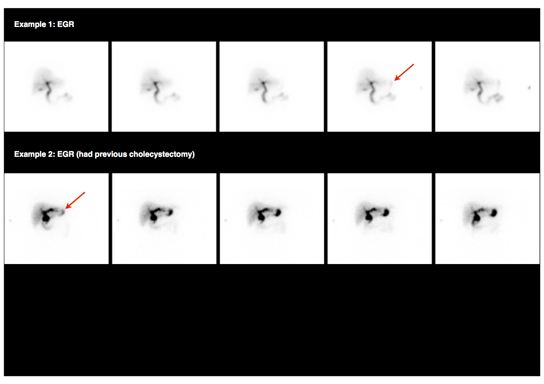





















Leave a Reply
You must be logged in to post a comment.