
A cancer with few visible symptoms
Ovarian cancer is often described as a “silent” disease because its early warning signs tend to be subtle and easy to overlook. Many of the initial symptoms are vague and can be attributed to common, noncancerous conditions, which makes early detection challenging. When reviewing Ovarian cancer images ultrasound findings, it becomes clear why the disease may go unnoticed in its beginning stages—there is usually no obvious external or visible indication.
In early-stage ovarian cancer, there is typically no outward visual proof of illness. Physical changes are rarely apparent without medical imaging, and this is why screening tools and symptom awareness are so important for women at risk.
Pictures
Symptoms of ovarian cancer
Early manifestations can include generalized abdominal discomfort, bloating, and a feeling of swelling in the lower abdomen. Some individuals report trouble eating or feeling full quickly after consuming only a small portion of food. Ovarian cancer may also trigger indigestion-like symptoms, pelvic or abdominal pain, and episodes of constipation, which are sometimes confused with irritable bowel syndrome.
As pressure from a growing tumor increases, you might notice a sudden urge to urinate or the need to urinate more frequently than usual. Certain women with ovarian cancer experience pain during intercourse. Changes in the menstrual cycle, such as irregular bleeding or altered flow, may also occur.
When the disease advances, symptoms generally become more persistent and pronounced. Ongoing fatigue, unintended weight loss, and unexplained back pain can all be associated with ovarian cancer. If symptoms are intense, progressively worsening, or last longer than a month, it’s important to consult your doctor promptly for evaluation and possible imaging, including Ovarian cancer images ultrasound assessment.
Abdominal swelling that does not resolve may raise further concern. For visual examples of how distension can appear, you can review Pictures of bloated stomach ovarian cancer. Many patients also wonder about symptom fluctuation, including Does ovarian cancer bloating go down, especially in early versus advanced stages.
Learn more: Ovarian cancer by the numbers: Facts, statistics, and you »
Diagnostic testing
Your physician will begin by reviewing your medical history and performing a comprehensive physical examination. The next step commonly involves a pelvic exam. While your primary care doctor may perform this evaluation, you could also be referred to a gynecologist for specialized assessment.
During a pelvic exam, a speculum is gently inserted into the vagina so the doctor can visually check for abnormalities. Using two gloved fingers inside the vagina while pressing on the abdomen with the other hand, the doctor assesses the uterus and ovaries for irregularities. Although this exam can provide useful clues, enlarged ovaries are not always detectable due to their deep position within the pelvis.
Blood tests
Your doctor will likely request blood work. One important marker is CA-125, a protein found on ovarian cancer cells that can be measured in the bloodstream. Elevated CA-125 levels may suggest ovarian cancer, but this result alone does not confirm a diagnosis. Blood tests may also help identify kidney or liver function abnormalities, which can influence treatment decisions.
Imaging tests
Imaging studies play a critical role in identifying structural abnormalities. They provide detailed information about the size, shape, and composition of the ovaries. While these tests can detect a mass or tumor, they cannot definitively determine whether it is cancerous. After a confirmed diagnosis, imaging is especially valuable in evaluating whether cancer has metastasized to other parts of the body.
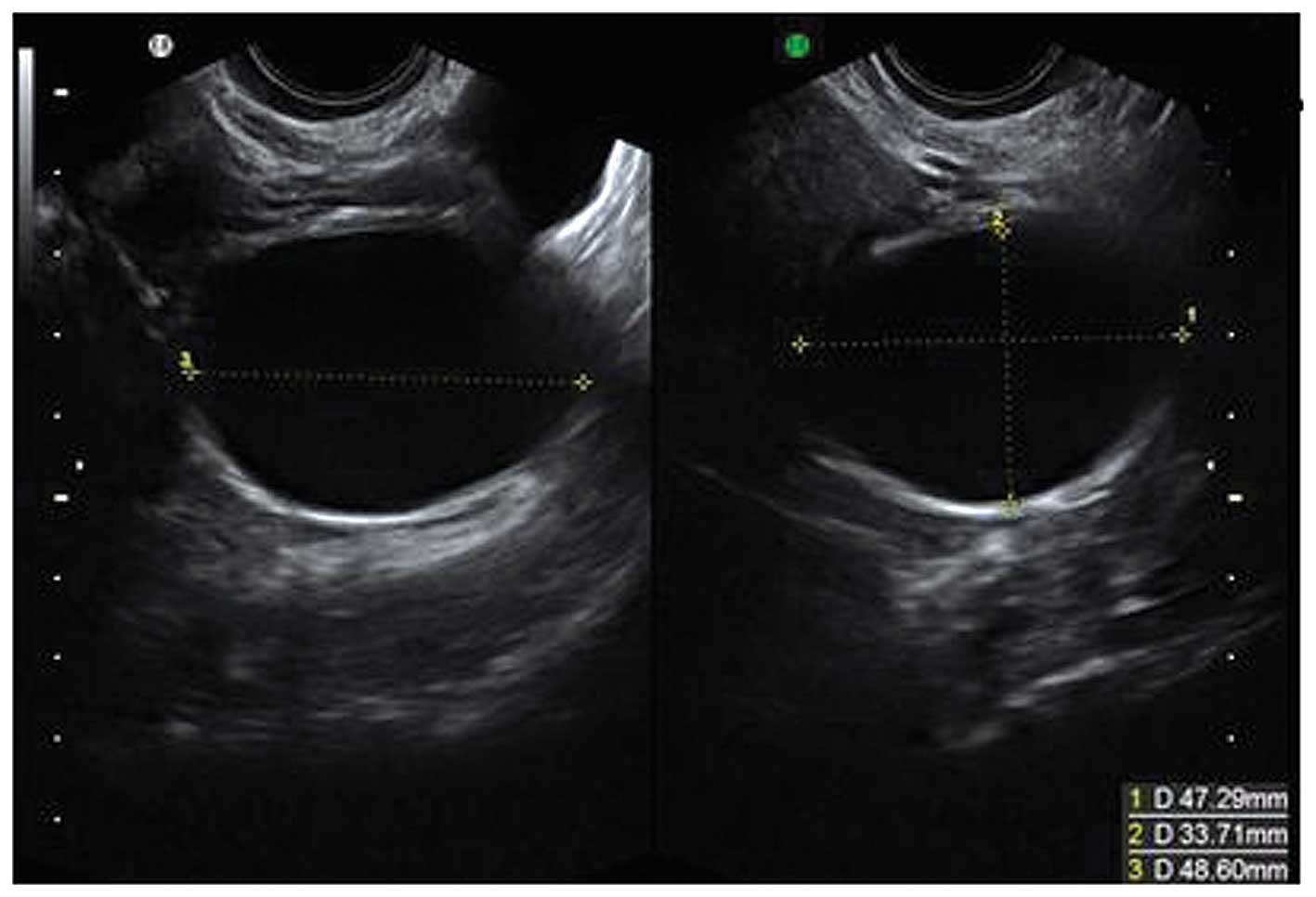
Ultrasound is often one of the first imaging tools used. During this procedure, a small probe is placed either on the abdomen or inside the vagina. Sound waves generate images that help visualize ovarian structures. High-quality Ovarian cancer images ultrasound scans can distinguish between solid tumors and fluid-filled cysts and may even reveal internal features of the ovaries that suggest malignancy.
Computed tomography (CT) scans use X-rays to produce cross-sectional images of the body. During the scan, you lie on a narrow table while the scanner rotates around you. An intravenous (IV) contrast dye may be administered to enhance image clarity.
Although CT scans may miss very small ovarian tumors, they are effective in identifying larger masses, swollen lymph nodes, and signs that cancer has spread beyond the ovaries.
MRI uses magnetic fields and radio waves to generate highly detailed images. It is frequently employed to obtain a clearer view of the ovaries and to further characterize masses initially seen on CT or ultrasound.
Chest X-rays may be performed to determine whether cancer has spread to the lungs. A positron emission tomography (PET) scan is not typically used as a primary diagnostic tool for ovarian cancer but can be helpful in detecting metastasis. PET scans utilize radioactive glucose to identify cancer cells based on their metabolic activity.
Surgery
In a minimally invasive procedure known as laparoscopy, your doctor inserts a thin, lighted tube through a small incision in the lower abdomen. This allows close inspection of the ovaries and nearby organs and tissues.
If ovarian cancer is suspected, the only definitive method to confirm the diagnosis is through biopsy. For ovarian cancer, this usually involves surgically removing the mass and sometimes one or both ovaries. A tissue sample is sent to a laboratory, where a pathologist examines it under a microscope. A pathologist is a physician trained to diagnose and classify diseases through microscopic evaluation. If fluid has accumulated in the abdomen, it can also be analyzed to determine whether cancer cells are present.
Risk factors for ovarian cancer
According to the Centers for Disease Control and Prevention (CDC), approximately 20,000 American women are diagnosed with ovarian cancer annually. The likelihood of developing ovarian cancer increases with age, and it is more common in middle-aged and older women.
Additional risk factors include:
- having a close relative with ovarian cancer, such as a mother, grandmother, sister, or aunt
- carrying the BRCA1 or BRCA2 gene mutation
- a personal history of breast, cervical, uterine, or colorectal cancer
- a previous diagnosis of melanoma or endometriosis
- having an Eastern European or Ashkenazi Jewish background
- never giving birth or experiencing fertility problems
- hormonal therapy — specifically, taking estrogen without progesterone for 10 years or more
If you have one or more of these risk factors and begin to notice symptoms associated with ovarian cancer, seek medical care without delay. Management may involve surgery, chemotherapy, radiation, or a combination of these treatments. Early detection through symptom recognition and appropriate imaging—such as Ovarian cancer images ultrasound—significantly improves prognosis and expands available treatment options.


Leave a Reply
You must be logged in to post a comment.