
Ever wondered why a sudden, fiery ache can strike a single joint out of nowhere, and why it sometimes feels like your body is fighting an invisible enemy? That sharp flare‑up is often crystal‑driven inflammation – a hidden but powerful trigger where tiny mineral crystals set off a cascade of immune fireworks. Within minutes, macrophages (the body’s “clean‑up crew”) spot these crystals, unleash inflammatory chemicals, and you’re left with swelling, heat, and pain that can mimic infection. Knowing how this works is the first step to stopping the fireworks before they burn out your joints for good.
How Crystals Wake Up the Immune System
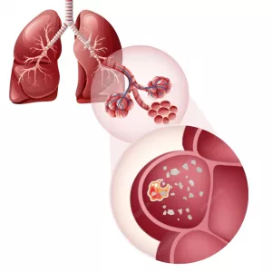
Crystals are not just boring little specks; they act like a rogue alarm button. When a crystal lands inside a joint or lung tissue, it’s taken up by macrophages. Inside the cell, the crystal can puncture the lysosome (the “trash can”), causing potassium to leak out, reactive oxygen species to rise, and the cell’s stress sensors to go off. This cascade lights up the NLRP3 inflammasome – a molecular “switch” that flips on and releases massive amounts of interleukin‑1β (IL‑1β) and IL‑18, the chemicals that bring on pain, redness, and swelling.
One recent study showed that the mechanosensitive channel TRPV4 is essential for this whole process. According to the TRPV4 research, blocking this channel dramatically reduces crystal‑induced inflammation in mouse models, highlighting a promising new target for therapy.
Crystal Types and Their Signature Diseases
Not all crystals are created equal. Different minerals form in different parts of the body, and each brings its own pattern of trouble.
Uric‑acid (MSU) crystals – the culprits of gout. They form when uric acid supersaturates the blood, often after a heavy meal or alcohol binge.
Calcium pyrophosphate (CPP) crystals – cause pseudogout, usually affecting the knee or wrist and often seen in older adults.
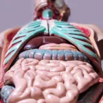
Cholesterol crystals – these appear in atherosclerotic plaques and can spill into circulation, igniting inflammation in blood vessels and occasionally in joints.
Silica and other dust particles – inhaled in mining or construction, they settle in the lungs, but the ensuing immune response can spill over into joints, leading to silicosis‑related arthritis.
Fat‑derived deoxysphingolipid crystals – a rare genetic defect that creates “fat crystals” which damage nerves, skin, and sometimes joints, as described in a recent ScienceDaily report.
All of these fall under the umbrella of inflammatory joint diseases, and understanding which crystal you’re dealing with guides the right treatment.
When Crystals Attack: Clinical Faces
Let’s walk through the most common ways crystal‑driven inflammation shows up in real life.
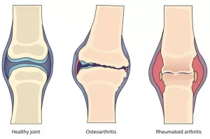
Gout and CPPD – The Classic Pair
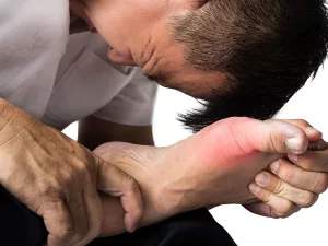
Picture this: You’re at a family dinner, indulge in a few too many shrimp tacos, and by morning your big toe is throbbing like a drum. That’s classic gout, driven by MSU crystals. The pain is excruciating, the joint is hot, and the skin may look shiny. Diagnosis is simple – pull a bit of synovial fluid and look under polarized light; you’ll see needle‑shaped, negatively birefringent crystals.
In pseudogout, the crystals are rhomboid and positively birefringent, often hitting the knee. The attacks are similar in intensity but the patient is usually older, and the crystals appear after a minor injury or surgery.
For a deeper dive into these twin conditions, check out our guide on gout and CPPD.
Silicosis‑Related Arthritis
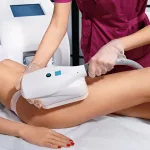
If you work with sand, stone, or concrete, you might breathe in silica dust. Over years, those particles lodge in the lungs, causing silicosis – a scarring disease that can also spark systemic inflammation. Some patients develop joint pain that mimics rheumatoid arthritis, but the underlying trigger is silica crystals activating macrophages far from the lungs.
Understanding the occupational link is crucial. Learn more about the root causes at our silicosis causes page.
Cholesterol‑Crystal Arthritis & Vascular Inflammation
When cholesterol builds up inside arteries, it can crystallize. These cholesterol crystals are not passive; they activate the NLRP3 inflammasome, just like urate crystals. The result? A burst of IL‑1β that can make joints painful and also fuel plaque instability. A study in the Journal of Immunology showed how cholesterol crystals directly trigger inflammasome activation, linking heart disease and joint pain.
Rare Fat‑Crystal Disorders
Some people inherit a defect that forces cells to produce deoxysphingolipids, which aggregate into crystal‑like lumps. These “fat crystals” clog mitochondria, especially in nerve cells, leading to pain, hearing loss, and skin lesions that heal poorly. While rare, the mechanism mirrors what we see with more common crystals – a physical obstruction that sparks inflammation.
Toxic‑Particle Diseases
Beyond silica, other occupational particles – asbestos, beryllium, even metal shavings – can become “toxic particles.” When macrophages ingest these, they launch the same inflammasome response, sometimes spilling over into joints. If you suspect this, read our article on toxic particle diseases for safety tips.
Finding the Culprit: Diagnosis Made Simple
Diagnosing crystal‑driven inflammation isn’t rocket science, but it does require a systematic approach.
Step 1 – Synovial‑Fluid Microscopy. Aspirate the joint, spread a drop on a slide, and examine under polarized light. The shape and birefringence tell you whether you’re looking at urate, CPP, or basic calcium phosphate crystals.
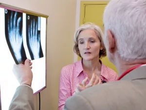
Step 2 – Imaging. Dual‑energy CT (DECT) can map urate deposits deep in tissue, while musculoskeletal ultrasound reveals the “double‑contour” sign of urate on cartilage. For silicosis‑related arthritis, a chest X‑ray or high‑resolution CT shows lung nodules that support the diagnosis.
Step 3 – Lab Panels. Check serum uric acid, calcium, phosphate, and lipid profile. Elevated CRP or ESR confirms inflammation but doesn’t pinpoint the crystal type.
Step 4 – Occupational History. Ask about dust exposure, construction work, or mining – the clues that point to silica or other toxic particles.
When the story is unclear, a careful review of inflammatory joint diseases can guide additional testing.
Calming the Fire: Management & Prevention
Now that we’ve identified the offender, how do we put it out?
Acute Relief
For most crystal attacks, start with NSAIDs (ibuprofen or naproxen) to quickly lower pain and swelling. If NSAIDs are contraindicated, colchicine is a classic alternative that blocks neutrophil activation. Short courses of oral corticosteroids (prednisone) work well when the flare is severe.
When IL‑1β is the main driver – as in gout or pseudogout – IL‑1 blockers (anakinra, canakinumab) can bring rapid relief. A review in Frontiers in Medicine noted that IL‑1 blockade resolves acute attacks within 24‑48 hours.
Long‑Term Strategies
Urate‑Lowering Therapy. All gout patients benefit from medications like allopurinol or febuxostat, which keep serum uric acid below 6 mg/dL, preventing crystal formation.
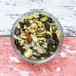
Diet & Lifestyle. Hydration is key – aim for at least 2‑3 liters of water daily. Limit high‑purine foods (organ meats, anchovies) and reduce alcohol, especially beer. For cholesterol‑crystal disease, adopt a Mediterranean diet low in saturated fats and rich in omega‑3 fatty acids.
Speaking of omega‑3, a 2018 study showed that omega‑3‑carboxylic acids dramatically reduced IL‑1β production in crystal‑stimulated macrophages (Nature Sci Reports), offering a dietary adjunct to medication.
Silica & Toxic Particles. The best prevention is avoidance. Use water‑based dust suppressants, wear respirators, and ensure proper ventilation on the job site. Once exposure has occurred, regular lung function testing can catch early disease before joint complications arise.
Targeting the Underlying Pathways
Scientists are exploring TRPV4 antagonists and NLRP3 inhibitors for future therapy. Early animal models suggest that blocking TRPV4 can blunt crystal‑induced IL‑1β release, while NLRP3 inhibitors (like MCC950) have shown promise in reducing gout flares in phase‑2 trials.
Follow‑Up & Monitoring
After an acute episode, schedule a follow‑up visit within 2‑4 weeks to reassess crystal load (via ultrasound or DECT) and adjust long‑term meds. Keep a symptom diary – note foods, alcohol, and activities that precede flares. This personal data becomes a powerful tool for you and your physician.
A Practical Checklist for Everyday Life
| Action | Why It Matters |
|---|---|
| Identify the crystal type | Directs specific treatment (e.g., urate‑lowering vs. silica avoidance) |
| Aspire joint fluid for microscopy | Gold‑standard diagnosis |
| Order targeted imaging (DECT, US) | Find hidden deposits |
| Start NSAID or colchicine promptly | Stops pain within hours |
| Consider IL‑1 blocker for severe flares | Targets the root cytokine |
| Adopt hydration & diet changes | Prevents new crystal formation |
| Use protective equipment at work | Reduces silica/toxic particle exposure |
| Track triggers in a diary | Empowers proactive management |
Conclusion
Crystal‑driven inflammation may feel like an unpredictable enemy, but it’s far from unbeatable. By understanding how tiny mineral shards hijack our immune system, recognizing the distinct diseases they cause, and applying a blend of quick‑acting meds, smart lifestyle tweaks, and emerging targeted therapies, you can keep those painful fireworks under control. Remember, the journey starts with a single step: getting the right diagnosis. From there, you have a clear roadmap to relief, fewer flares, and healthier joints for the long haul. If you have questions about your own symptoms or want personalized advice, reach out to a trusted rheumatologist—your future self will thank you.


Leave a Reply
You must be logged in to post a comment.