Hey there! If you’ve ever heard the term “intraocular pressure” and thought, “Sounds like something only doctors need to worry about,” you’re not alone. The truth is, this little number can tell us a lot about the health of our eyes—sometimes even before we notice any change in vision. Think of it like the tire pressure on a bike: keep it just right, and the ride is smooth; too high or too low, and you’re headed for a wobble.
In the next few minutes, I’m going to walk you through the basics, why we care about high eye pressure, how it’s measured, and what you can actually do to keep it in check. Grab a cup of tea, settle in, and let’s chat about one of the most important “vital signs” for your eyes.
Understanding IOP Basics
What is intraocular pressure?
Intraocular pressure (IOP) is the fluid pressure inside the eye. It’s created by the constant flow of a clear liquid called aqueous humor, which is produced by the ciliary body, circulates around the front of the eye, and then drains out through a tiny meshwork called the trabecular meshwork. When production and outflow are balanced, the pressure stays within a healthy window.
Normal intraocular pressure range
Most healthy eyes sit comfortably between 10 and 21 mm Hg. However, the “normal” range can shift a bit during the day and from person to person. Below is a quick reference table you can keep on your fridge or phone.
| Age Group | Typical IOP (mm Hg) |
|---|---|
| Children (0‑12) | 12‑16 |
| Teens (13‑19) | 13‑18 |
| Adults (20‑40) | 12‑20 |
| Adults (40+) | 13‑21 |
Why IOP matters
IOP isn’t just a number; it’s the only modifiable risk factor for glaucoma, the “silent thief of sight.” When pressure is too high, it can compress the optic nerve, gradually stealing peripheral vision without the person noticing until the damage is already done. Keeping the pressure in a safe range helps maintain the eye’s shape, nutrient flow, and overall visual clarity.
How eye pressure is measured
Eye‑care professionals use a variety of tonometry devices to gauge IOP. The gold‑standard is Goldmann applanation tonometry, which gently flattens a tiny area of the cornea and measures the force required. For quick screenings, many offices use a non‑contact “air‑puff” tonometer that feels like a brief puff of air on the eye.
Other handy tools include handheld devices such as the Tono‑Pen and iCare rebound tonometer—great for kids, people with corneal irregularities, or community screenings. EyeWiki provides a full rundown of these methods, and you’ll soon see why your eye doctor might choose one over another.
When Pressure Gets High
What qualifies as “high” IOP?
Anything consistently above 21 mm Hg is considered elevated. The higher the number, the greater the risk—especially if readings climb past 27 mm Hg, where optic‑nerve damage becomes more likely.
Ocular hypertension vs. glaucoma
“Ocular hypertension” simply means the pressure is high but there’s no detectable damage to the optic nerve or visual field yet. Some people live with pressures of 25 mm Hg their whole lives without developing glaucoma; others may experience optic‑nerve loss even with pressures that look “normal.” This is why regular monitoring is essential.
Risks of untreated high eye pressure
Besides glaucoma, sustained high IOP can lead to:
- Progressive optic‑nerve damage
- Reduced peripheral vision (often described as “tunnel vision”)
- Potentially irreversible vision loss if left unchecked
According to Optometrists.org, glaucoma is responsible for vision loss in over 12 % of the population worldwide—so keeping an eye on the pressure is a proactive step we can all take.
When to get re‑checked
If you’ve been diagnosed with ocular hypertension, your doctor will likely schedule follow‑up visits every 3‑6 months, adjusting the frequency based on how stable your numbers are. Even if you never had a high reading, it’s smart to have a baseline check at age 40 and then every 1‑2 years after that.
How IOP Is Measured
Goldmann Applanation Tonometry
This slit‑lamp‑mounted device is the clinical “gold standard.” A tiny probe gently flattens the cornea while a fluorescein dye highlights the contact area. The measured force directly translates to IOP in mm Hg. It’s highly accurate but requires a cooperative patient and a skilled examiner.
Non‑Contact “Air‑Puff” Tonometry
Quick, painless, and doesn’t need any eye drops. A brief puff of air flattens the cornea just enough for the device to gauge pressure. Perfect for routine screenings, though it can be a little startling the first time!
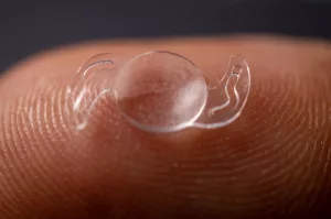
Handheld Devices (Tono‑Pen, iCare)
These portable tonometers are great for children, elderly patients, or remote clinics. They use a small probe that briefly touches the cornea, delivering a quick reading without anesthesia. Their convenience comes with slightly more variability, so they’re often used as a supplement to, not a replacement for, Goldmann measurements.
Dynamic‑Contour Tonometry
Unlike applanation, this device measures pressure without flattening the cornea, reducing errors from corneal thickness. It’s especially useful for patients with unusually thin or thick corneas.
Continuous IOP Monitoring
Emerging technologies now allow real‑time pressure tracking via tiny, implantable sensors. While still largely in research phases, they promise a future where sudden spikes can be caught before they cause damage.
Complementary glaucoma tests
IOP is just one piece of the puzzle. Your eye doctor will likely also order visual‑field tests, optical‑coherence tomography (OCT) of the optic nerve, and pachymetry to measure corneal thickness—all of which help interpret the pressure reading in context.
Ways to Lower IOP
Prescription eye drops
The most common first‑line treatment. There are several classes:
- Prostaglandin analogs (e.g., latanoprost) – boost outflow.
- Beta‑blockers (e.g., timolol) – reduce production.
- Carbonic anhydrase inhibitors (e.g., dorzolamide) – also cut production.
- Alpha‑agonists and Rho‑kinase inhibitors – work in various pathways.
Following the drop schedule precisely is crucial. A simple tip: place the drop, close your eye gently for a minute, and press your inner corner to prevent drainage into the nose.
Laser therapies
Selective laser trabeculoplasty (SLT) and argon laser trabeculoplasty (ALT) use tiny laser pulses to improve fluid outflow through the trabecular meshwork. Many patients achieve a pressure drop of 20‑30 % without medication, at least temporarily.
Surgical options
When drops and lasers aren’t enough, surgeons may perform a trabeculectomy (creating a new drainage pathway) or implant tiny shunts. Minimally invasive glaucoma surgery (MIGS) devices are gaining popularity for their quicker recovery and lower complication rates.
Lifestyle modifications
While lifestyle changes alone rarely normalize a high IOP, they can complement medical therapy:
- Diet: leafy greens (lutein, zeaxanthin), omega‑3‑rich fish, and vitamin‑rich foods support ocular health.
- Exercise: moderate cardio (walking, cycling) has been shown to modestly lower IOP.
- Caffeine: limit excess—high doses may cause short‑term spikes.
- Hydration: drink water steadily throughout the day; large sudden volumes can raise pressure temporarily.
- Sleep position: elevating the head with an extra pillow can reduce overnight pressure.
These habits are modest but can tip the balance in your favor, especially when paired with proper medication.
Adherence & monitoring
Even the best regimen fails if it isn’t used consistently. Set reminders on your phone, keep a medication diary, and schedule regular follow‑ups. Some patients find home‑tonometry devices (like iCare HOME) handy for tracking trends between office visits.
Quick FAQ Answers
What is a normal intraocular pressure?
Typically 10‑21 mm Hg, though individual “normal” can shift slightly.
How is eye pressure measured?
Using a tonometer—Goldmann applanation is the clinical gold standard, while air‑puff and handheld devices are common for quick checks.
Can glaucoma occur with normal IOP?
Yes—about a third of glaucoma cases are “normal‑tension glaucoma,” where optic‑nerve damage happens despite pressure within the normal range.
What does ocular hypertension mean?
Pressure above 21 mm Hg without detectable optic‑nerve damage. It warrants close observation.
How often should I get my IOP checked?
At least once every 1‑2 years after age 40, or more often if you have risk factors or are on treatment.
Are there home IOP devices?
Yes—handheld rebound tonometers like iCare HOME let you monitor pressure under a doctor’s guidance.
Expert Insights & Sources
To keep this guide trustworthy, I consulted a range of reputable sources:
- StatPearls’s detailed physiology chapter on IOP (NCBI).
- The American Academy of Ophthalmology’s EyeWiki overview of tonometry (EyeWiki).
- Clinical recommendations from the Cleveland Clinic (Cleveland Clinic).
- Practical lifestyle advice from the Glaucoma Research Foundation (Glaucoma.org).
These sources are peer‑reviewed, up‑to‑date, and written by ophthalmologists, ensuring the information you’re reading is both accurate and actionable.
Takeaway Cheat Sheet
Key numbers: Normal IOP = 10‑21 mm Hg; High = > 21 mm Hg; Risk climbs sharply > 27 mm Hg.
When to act: Any persistent rise, optic‑nerve changes, or visual‑field loss.
Top three ways to manage: Prescription drops, laser therapy, healthy lifestyle.
Quick check list for appointments:
- Ask about your latest IOP reading.
- Discuss any side‑effects from drops.
- Review your family history of glaucoma.
- Confirm next follow‑up date.
Remember, intraocular pressure is just one piece of the eye‑health puzzle, but it’s a piece you can actively influence. By staying informed, keeping up with regular eye exams, and joining forces with your eye doctor, you give your vision the best chance to stay bright for years to come.
Got a question about your own eye pressure? Or perhaps a story about how you tackled high IOP? Share it in the comments below—I’d love to hear from you! And if this guide helped clear the fog, feel free to pass it along to a friend who might need a gentle reminder that their eyes deserve the same care we give any other part of our bodies.


Leave a Reply
You must be logged in to post a comment.