Ever wondered how scientists can actually see the tangled web of wires inside your skull? In just a few sentences: 3D brain connections are high‑resolution, three‑dimensional maps that show every major nerve fiber pathway and how they talk to each other. These maps are the newest language we have for talking about the brain’s “connectome,” and they’re already reshaping everything from basic research to the operating room.
What’s even cooler is that today you don’t need a PhD in neurobiology to peek at these maps. Free online portals let you spin, zoom, and explore the same data that Harvard‑Google teams used to build the most detailed wiring diagram of a human brain ever made. In the next few minutes we’ll break down what 3D brain connections are, why they matter, the biggest projects behind them, and how you can start exploring them right now – all in plain‑English, with a few stories and jokes along the way.
What Are 3D Connections
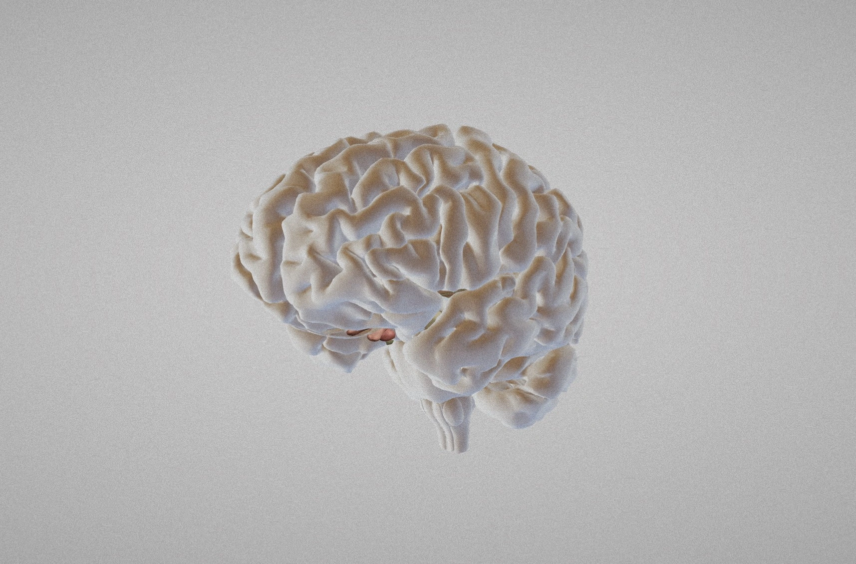
At its core, a 3D brain connection map (or “connectome”) is a digital reconstruction that captures the shape, direction, and length of nerve fiber pathways throughout the entire brain volume. Think of it like a city’s subway map, but instead of stations and lines you have neurons and axons, and the map is built in three dimensions so you can fly through it from any angle.
Traditional brain imaging gave us flat, two‑dimensional slices – kind of like looking at a pizza from the side. 3D techniques stack those slices and fill in the gaps, creating a full‑blown, volumetric model you can walk around in a virtual reality headset or on a regular computer screen. The result is a window into the brain’s wiring that’s both detailed enough for scientists and, thanks to modern tools, increasingly accessible to curious minds.
Key terms you’ll hear a lot:
- Brain neuroimaging – the suite of methods (MRI, CT, PET, etc.) that let us see the living brain.
- Nerve fiber pathways – bundles of axons that transmit signals between different brain regions.
- Nerve fiber imaging – techniques specifically tuned to capture those pathways, often using diffusion‑weighted MRI.
- Neuroimaging techniques – a broader category that includes everything from functional MRI to electron microscopy.
For a beginner‑friendly dive into the basics of brain neuroimaging, check out our quick guide – it’s a great primer before you start fiddling with 3D data.
Why It Matters
Seeing is believing, and when you can actually visualise how neurons are wired, a whole new world of possibilities opens up.
Clinical Benefits
Imagine a neurosurgeon planning to remove a tumor that sits right next to language‑processing areas. With a 3D tractography map, they can plot the exact route of the language pathways and steer clear of them, preserving the patient’s ability to speak. That’s not sci‑fi; it’s already happening in major hospitals worldwide.
3D maps also help catch neurodegenerative diseases early. Patterns of white‑matter loss can be spotted before symptoms become obvious, giving doctors a head start on treatments for conditions like Alzheimer’s or multiple sclerosis.
Research Power
Researchers can finally ask “big picture” questions: How many connections does a typical human cortex have? Are certain pathways more susceptible to stress? The answer to those queries is the kind of data that was once a dream, now a reality thanks to projects like the Human Connectome Project (HCP) and the Allen Institute’s Mouse Brain Connectivity Atlas.
And there’s a flip side: every new map also reveals gaps in our knowledge that spark fresh investigations. The field is moving fast, and every new connection you see is a potential clue about how thoughts, memories, and emotions emerge.
Potential Risks & Ethics
With great power comes great responsibility. High‑resolution brain maps contain intimate details about an individual’s neural architecture, and that data could be misused if privacy isn’t protected. Also, the public sometimes jumps to conclusions—seeing a connection on a map doesn’t automatically mean it explains a behavior or disease. That’s why scientists keep a cautious tone and why we, as readers, should stay critical.
According to the NIH Human Connectome Project[1], robust data‑sharing policies and de‑identification steps are in place to curb these risks. Still, awareness is key, and conversations about ethics should go hand‑in‑hand with technological advances.
Leading Projects
Harvard‑Google 3D Connectome
In 2024 Wired ran a story about a collaborative effort that built a 3‑dimensional wiring diagram from a single cubic millimeter of a woman’s cortex. The map contains roughly 57,000 cells and 150 million synapses—imagine a city with millions of streets, each instantly traceable. The team combined electron microscopy (to capture nanometer‑scale detail) with machine‑learning algorithms that stitched the images together into a navigable 3‑D space.
As reported by Wired[2], this level of detail lets scientists test “wiring rules” that could explain how thoughts form or why certain disorders arise.
Allen Institute – Mouse Brain Connectivity Atlas & MICrONS Explorer
The Allen Institute offers a free, web‑based viewer that lets you explore mouse brain pathways across thousands of brain regions. It’s an excellent sandbox for anyone new to 3D brain maps because the interface is intuitive, and the data are openly downloadable.
Learn more about the platform here.
BigBrain – Ultra‑High‑Resolution Human Model
BigBrain is a massive 1‑terabyte dataset that reconstructs a whole human brain at 20‑micrometer resolution—close to the size of a single neuron. It bridges the gap between macro‑scale MRI and micro‑scale histology, letting researchers zoom from whole‑brain anatomy down to individual cortical layers.
Details on the project are available from the McGill Centre for Integrative Neuroscience[4].
Explore Yourself
Feeling inspired? You don’t need a lab coat to start exploring 3D brain connections. Here’s a simple, step‑by‑step guide that anyone can follow.
Step 1 – Pick a Viewer
If you like sleek, browser‑based tools, try Neuroglancer (the Allen Institute’s viewer). For a more hands‑on experience, download a sample dataset from the HCP and open it in the free software FSLNets – it creates connectivity matrices you can play with.
Step 2 – Get Sample Data
Head over to the HCP data portal and grab a small tractography set (a few megabytes is enough for a first look). These files are usually in NIfTI format, which most viewers accept out of the box.
Step 3 – Load & Visualise
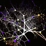
Drag the file into your chosen viewer. You’ll see a cloud of colorful lines dancing through a 3‑D brain silhouette. Toggle the opacity, change the color map, or isolate a single pathway to see how it weaves from the frontal lobe to the occipital lobe.
Step 4 – Play with Metrics
Most tools let you compute basic stats: fiber count, average length, and density per region. These numbers can hint at which pathways are most “busy” in a given brain – a fun way to practice data‑driven storytelling.
Along the way, you might want to learn more about how these pathways are classified, so feel free to visit our page on nerve fiber pathways for a quick taxonomy.
Tools Checklist
| Tool | Purpose | Key Feature |
|---|---|---|
| Neuroglancer | 3‑D visualization | Web‑based, GPU‑accelerated |
| FSLNets | Connectivity matrices | Statistical analysis of networks |
| MATLAB / Python (Nilearn) | Custom scripts | Open‑source, flexible |
| BigBrain Viewer | Ultra‑high‑resolution human data | 20‑µm voxel size |
Troubleshooting Tips
- Make sure your data are in NIfTI (.nii) format; if not, use
dcm2niixto convert. - If the viewer runs slowly, lower the resolution or turn off unnecessary overlays.
- For GPU‑heavy visualizations, ensure your browser supports WebGL.
Need a deeper dive into the nuts and bolts of the imaging methods themselves? Our guide on neuroimaging techniques walks you through diffusion MRI, tractography, and even emerging expansion microscopy.
Future Directions
The road ahead looks exhilarating. Here are a few trends to watch.
Emerging Technologies
Expansion microscopy—where tissue is physically enlarged before imaging—promises even finer detail without the need for massive electron microscopes. Combined with AI‑driven segmentation, we could soon map whole human brains at synapse‑level resolution in a matter of weeks instead of years.
Large‑Scale Initiatives
Beyond the HCP, the European Human Brain Project and the US BRAIN Initiative are racing to create an “atlas of everything” – a seamless integration of genetics, electrophysiology, and 3D connectivity. Their ultimate goal? A digital twin of the human brain that can predict how a specific drug will affect a patient’s network dynamics.
Personalized Medicine
Imagine a future where your doctor orders a quick diffusion‑MRI scan, feeds the data into a cloud‑based algorithm, and gets a personalized risk profile for dementia based on subtle changes in your own white‑matter pathways. That vision is already being prototyped in a few cutting‑edge clinics.
Wrapping Up
We’ve travelled from the basic idea of “what is a 3D brain connection?” to the cutting‑edge maps that Harvard and Google built, and even walked through how you can explore these marvels on your own laptop. The big takeaway? 3D brain connections are not just abstract scientific curiosities; they’re concrete tools reshaping diagnosis, treatment, and our fundamental understanding of the mind.
If you’re curious to keep learning, start by playing with a free dataset, read up on nerve fiber imaging, and stay tuned to the latest releases from the Human Connectome Project. The brain is the ultimate puzzle, and every new map you explore is a piece of that puzzle falling into place.
What part of the brain’s wiring are you most excited to discover? Drop a thought, share your own adventure with a 3D viewer, or ask any question that pops up while you’re exploring. Let’s keep the conversation going – after all, the best discoveries happen when we share them together.


Leave a Reply
You must be logged in to post a comment.