Imagine your brain as a bustling city. The amygdala is the neighborhood watch – a compact but critical district that decides whether the traffic lights turn green for fight, flight, or a friendly wave. The tiny workers that run this watch are the amygdala neurons. In the next few minutes you’ll meet the different crews, learn why they matter to your everyday mood, and see how the newest science could one day help you feel steadier.
Ready to explore? Let’s dive in together and see how a handful of cells shape everything from a sudden startle to the warm glow when you see a familiar face.
What Are Amygdala Neurons?
Definition and basic anatomy
The amygdala sits deep inside the medial temporal lobe, nestled next to the hippocampus. It’s not a single structure but a collection of nuclei – the lateral (LA), basolateral (BLA), central (CeA), and medial (MeA) among others. Each nucleus hosts its own families of neurons, all wired to read, tag, and broadcast emotional value.
How they differ from nearby hippocampus or visual‑feature cells
While hippocampus neurons specialize in stitching together the timeline of an experience, amygdala neurons slap an emotional “stamp” onto that timeline. Visual‑features recognition pathways first tell the brain “there’s a red apple,” and the amygdala decides whether that apple is delicious, dangerous, or somewhere in between.
Quick fact box
- ≈ 1 million neurons in the human amygdala.
- Only ~5 % are GABA‑ergic interneurons, yet they regulate >70 % of output firing.
- Value‑coding neurons can predict a choice with >90 % accuracy (Grabenhorst et al., 2013).
Major Types Explained
Excitatory projection neurons (glutamatergic)
These are the workhorses that send messages out of the amygdala. In the basolateral complex, pyramidal neurons fire glutamate onto the central nucleus, the prefrontal cortex, and the hippocampus. Their activity is the heartbeat of fear learning – they light up when you hear a startling sound and keep the signal alive long enough for a memory to form.
Inhibitory interneurons (GABA‑ergic) – the “gatekeepers”
Even though they’re a minority, GABA‑ergic cells are the traffic controllers. Parvalbumin‑positive (PV) cells act like stoplights, rapidly silencing excessive firing. Somatostatin‑positive (SST) cells provide a slower, “brake‑by‑curve” inhibition that shapes the timing of fear responses. VIP‑expressing cells modulate the other interneurons, allowing for a nuanced balance between excitation and inhibition.
Specialized sub‑populations
Research on monkeys doing a juice‑choice task revealed a group of amygdala neurons that switch from “value coding” (how good the juice is) to “choice coding” (which lever you’ll press). When the same neurons were silenced, the animals’ predictions fell apart, showing these cells are essential for decision making.
Anxiety‑selective neurons
A 2011 study found a specific set of basolateral cells that fire tonically only when an animal feels anxious in an open field. Their ramp‑up pattern mirrors how your heart slowly speeds up while you wait for a test result.
Neuron type | Neurotransmitter | Primary outputs | Key behavior
| Neuron type | Neurotransmitter | Main targets | Representative behavior |
|---|---|---|---|
| Lateral‑BLA pyramidal | Glutamate | CeA, prefrontal cortex, hippocampus | Fear acquisition |
| PV‑interneuron | GABA | Local pyramidal cells | Fast inhibition, timing |
| ITC cluster (intercalated) | GABA | BLA & CeA | Threat‑imminence modulation |
| Anxiety‑selective BLA cell | Glutamate | Nucleus accumbens, ventral hippocampus | Gradual anxiety rise |
Neurons in Daily Life
Fear & anxiety processing
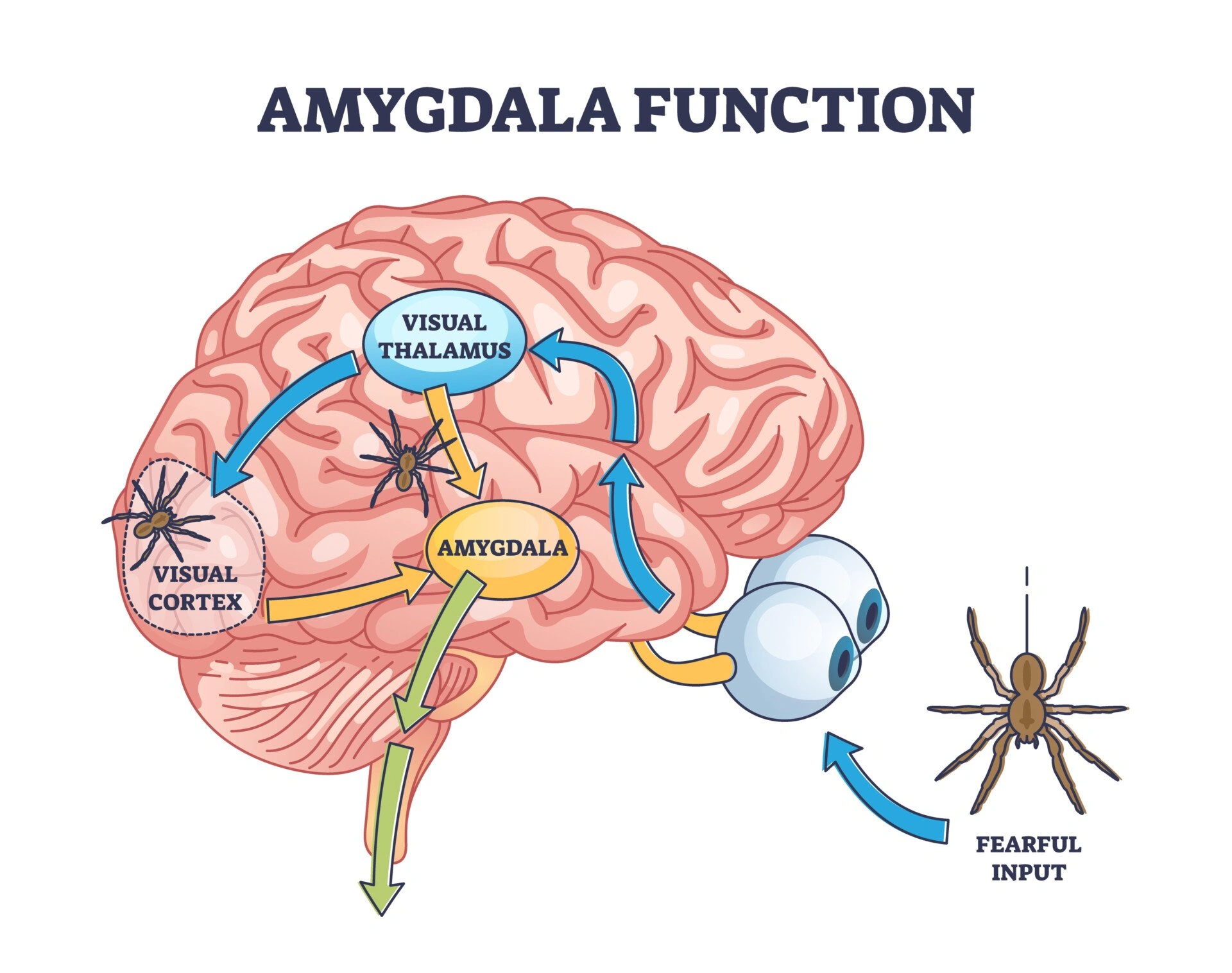
When a sudden car horn blares, sound information travels from the thalamus to the lateral amygdala. Within milliseconds, excitatory neurons ignite, and the central nucleus launches a cascade that speeds up your heart and tenses muscles. That’s why you can feel a shiver even before you consciously recognize the danger.
Social connection & face‑recognition neurons
Seeing a smiling friend isn’t just a visual thing. Face‑recognition neurons in the temporal cortex send a “this person is safe” signal to the amygdala, which then tags the encounter with positive valence. When that tag is missing – as can happen in some forms of autism – social cues feel flat or ambiguous.
Memory linking – amygdala ↔ hippocampus
The hippocampus writes the story; the amygdala adds the emotional color. Studies show that silencing amygdala output during learning makes memories weaker and less vivid. It’s why the taste of a dish you ate on a bad day can stay stuck in your mind forever.
Mini‑case study
Imagine you’re doing a visual features recognition test while a faint alarm buzzes in the background. fMRI scans reveal heightened activity in both the lateral amygdala and the visual cortex. The amygdala’s “alert” signal boosts attention to the visual task – a protective mechanism that ensures you don’t miss important details when something might be dangerous.
Research Methods & Findings
Electrophysiology & single‑cell recordings
Microelectrodes placed in awake monkeys have captured how amygdala neurons transition from valuing a reward to committing to a choice. The data show a gradual ramp‑up of firing rates that mirrors the animal’s internal deliberation.
Single‑cell RNA‑seq atlases (2023)
Using single‑cell transcriptomics, scientists identified 130 distinct neuronal sub‑types in the adult mouse amygdala. A subset of these cells turned on genes for neurite outgrowth and synaptic plasticity after fear conditioning – a molecular glimpse of the “engram” (memory trace).
Optogenetics & chemogenetics – manipulating specific populations
When researchers shine light on intercalated cells (ITCs), the animals’ defensive behavior flips between passive freezing and active escape. This demonstrates that ITCs act like a central switchboard, balancing competing survival strategies.
Expert insight
Dr. Limeng Huang, a neuro‑biologist at Zhejiang University, says, “Understanding the input‑output organization of amygdalar circuits lets us predict how a single neuron’s activity ripples across the brain.” His work underscores why precise control of intercalated cells is a hot target for future anxiety treatments.
Clinical Relevance Today
Anxiety disorders & PTSD
Hyper‑active anxiety‑selective BLA neurons can cause over‑generalized fear, a hallmark of PTSD. Pharmacological agents that boost GABAergic tone are already helping patients, but the next wave may involve gene‑editing tools that silence the over‑active ensembles identified in the RNA‑seq atlas.
Depression & social‑cognition deficits
When the amygdala’s communication with face‑recognition circuits falters, social cues lose their emotional weight. This can contribute to the social withdrawal seen in major depressive disorder. Therapies that restore balanced amygdala‑cortical connectivity are being explored in clinical trials.
Potential therapeutic targets
Modulating ITC clusters, fine‑tuning PV interneurons, or delivering viral vectors that adjust activity‑responsive genes are all on the research horizon. The goal? A “smart” treatment that dampens pathological fear without dulling normal emotional responsiveness.
Practical checklist for everyday life
- Notice early signs of an over‑active amygdala: rapid heartbeat, racing thoughts, or exaggerated startle.
- Practice mindfulness or slow breathing – both increase GABAergic inhibition and can calm the circuitry.
- Maintain regular aerobic exercise; studies link it to healthier amygdala‑prefrontal connectivity.
- Prioritize good sleep – the brain consolidates the emotional tags during REM, reducing next‑day anxiety.
Conclusion
Amygdala neurons are the brain’s rapid‑response team, turning raw sensory flashes into the rich tapestry of feelings that guide us every day. By distinguishing the excitatory “broadcast” cells from the inhibitory “gatekeepers,” and by appreciating the newest molecular maps that reveal over a hundred sub‑types, we begin to understand why a fleeting threat can linger as a scar, or why a familiar smile can instantly lift our spirit.
Beyond satisfying curiosity, this knowledge lights the path toward smarter treatments for anxiety, PTSD, and social‑cognition challenges. As optogenetics, single‑cell genomics, and AI‑driven modeling converge, the next decade may let us gently retune amygdala neurons for healthier emotional lives.
What part of the amygdala’s story surprised you the most? If you have questions or want to share a personal experience about how emotions shape your daily decisions, feel free to reach out. We’re all in this neural adventure together.


Leave a Reply
You must be logged in to post a comment.