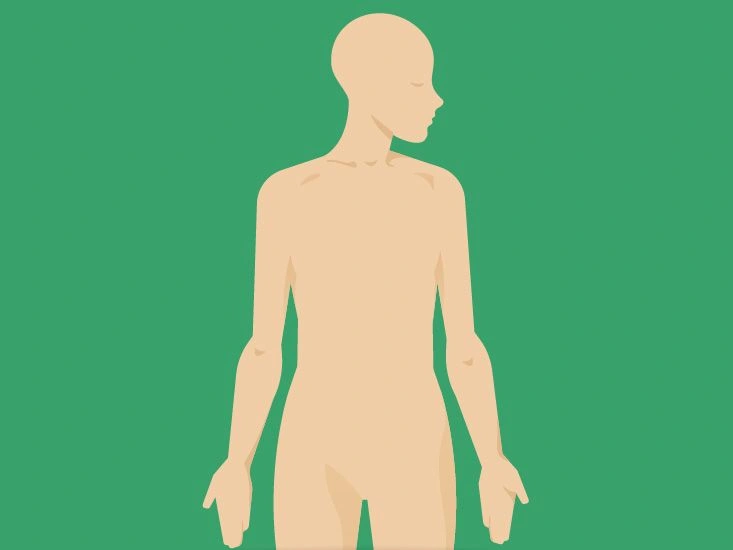
The arms are the body’s upper limbs. They rank among the most intricate and frequently used regions of the body.
Each arm is made up of four primary sections:
- upper arm
- forearm
- wrist
- hand
Continue reading to discover more about the bones, muscles, nerves, and vessels of the upper arm and forearm, along with common arm conditions you might encounter.
Anatomy
Upper arm
The upper arm encompasses the shoulder and the region between the shoulder and the elbow joint. The main bones of the upper arm are:
- Scapula. Also known as the shoulder blade, the scapula is a triangular, flat bone mainly anchored by muscles. It connects the arm to the trunk.
- Clavicle. The clavicle, or collarbone, also anchors the arm to the torso and helps transfer force from the upper limb to the skeleton.
- Humerus. The humerus is the long bone of the upper arm situated between the scapula and the elbow. Numerous muscles and ligaments attach to the humerus.
The upper arm includes several joints as well, including the:
- Acromioclavicular joint. Where the scapula meets the clavicle.
- Glenohumeral joint. The joint between the scapula and humerus.
- Sternoclavicular joint. Where the clavicle meets the sternum (breastbone).
Forearm
The forearm lies between the elbow and the wrist. It contains two primary bones: the radius and the ulna.
- Radius. Positioned on the thumb side of the forearm, the radius rotates around the ulna and alters its orientation depending on hand movement. Many muscles attach to the radius to facilitate movement at the elbow, wrist, and fingers.
- Ulna. Running parallel to the radius, the ulna sits on the side nearer the little finger. Unlike the radius, the ulna remains relatively fixed and does not rotate.
Elbow joint
The elbow joint is where the humerus of the upper arm meets the radius and ulna of the forearm.
The elbow is actually composed of three linked joints:
- Ulnohumeral joint. The connection between the humerus and the ulna.
- Radiocapitellar joint. Where the radius articulates with the capitellum region of the humerus.
- Proximal radioulnar joint. The joint linking the radius and ulna, enabling rotation of the hand.
Anatomy and function of upper arm muscles
The upper arm is divided into two compartments: the anterior compartment and the posterior compartment.
Muscle movement
Before reviewing the specific muscles, it helps to understand four primary types of movement these muscles perform:
- Flexion. Movement that brings two parts closer together, such as bending the elbow so the forearm approaches the upper arm.
- Extension. Movement that increases the distance between two parts, for example straightening the elbow.
- Abduction. Moving a part away from the body’s midline, like raising the arm out to the side.
- Adduction. Moving a part toward the midline, for instance bringing the arm back to rest alongside the torso.
Anterior compartment
Located in front of the humerus, the anterior compartment contains these muscles:
- Biceps brachii. Commonly called the biceps, this muscle has two heads that originate near the shoulder and merge near the elbow. Its distal portion flexes the forearm, drawing it toward the upper arm. The proximal heads assist with flexion and adduction of the upper arm.
- Brachialis. Situated beneath the biceps, the brachialis links the humerus to the ulna and is a primary flexor of the forearm.
- Coracobrachialis. Located close to the shoulder, this muscle permits adduction and flexion of the upper arm and helps stabilize the humerus in the shoulder socket.
Posterior compartment
Found behind the humerus, the posterior compartment comprises two muscles:
- Triceps brachii. Usually called the triceps, this muscle runs along the humerus and is responsible for extension of the forearm; it also contributes to shoulder stabilization.
- Anconeus. A small triangular muscle that aids elbow extension and forearm rotation, sometimes viewed as an extension of the triceps.
Anatomy and function of forearm muscles
The forearm contains a greater number of muscles than the upper arm. It’s divided into anterior and posterior compartments, each further organized into layers.
Anterior compartment
Running along the inner aspect of the forearm, the anterior compartment primarily enables flexion of the wrist and fingers, and rotation of the forearm.
Superficial layer
- Flexor carpi ulnaris. Flexes and adducts the wrist.
- Palmaris longus. Assists wrist flexion; absent in some individuals.
- Flexor carpi radialis. Facilitates wrist flexion and abduction of the hand and wrist.
- Pronator teres. Rotates the forearm so the palm faces inward.
Intermediate layer
- Flexor digitorum superficialis. Flexes the second through fifth fingers.
Deep compartment
- Flexor digitorum profundus. Also flexes the fingers and assists in drawing the wrist toward the body.
- Flexor pollicis longus. Flexes the thumb.
- Pronator quadratus. Works with pronator teres to rotate the forearm.
Posterior compartment
Located along the back of the forearm, the posterior compartment contains muscles that extend the wrist and fingers.
Unlike the anterior compartment, it lacks an intermediate layer.
Superficial layer
- Brachioradialis. Flexes the forearm at the elbow.
- Extensor carpi radialis longus. Aids in wrist abduction and extension.
- Extensor carpi radialis brevis. The shorter, broader partner to the longus muscle.
- Extensor digitorum. Extends the second through fifth fingers.
- Extensor carpi ulnaris. Adducts the wrist.
Deep layer
- Supinator. Rotates the forearm outward so the palm faces up.
- Abductor pollicis longus. Moves the thumb away from the hand.
- Extensor pollicis brevis. Extends the thumb.
- Extensor pollicis longus. The longer companion muscle that also extends the thumb.
- Extensor indicis. Extends the index finger.
Brachial plexus
The brachial plexus is a network of nerves that supply the skin and muscles of the arm. It originates from the spinal column and extends down the arm.
The brachial plexus is organized into five segments:
- Roots. The plexus begins with roots formed by the spinal nerves C5, C6, C7, C8, and T1.
- Trunks. Three trunks arise from the roots: superior (C5–C6), middle (C7), and inferior (C8–T1).
- Divisions. Each trunk splits into anterior and posterior divisions, making six divisions total.
- Cords. The anterior and posterior divisions recombine to form three cords named lateral, posterior, and medial.
- Branches. The cords give rise to the peripheral nerves that innervate the arm.

Peripheral nerves
Peripheral nerves in the arm handle both motor control and sensation.
Key peripheral nerves of the arm include:
- Axillary nerve. Passes between the scapula and humerus, innervating shoulder muscles such as the deltoid, teres minor, and a portion of the triceps.
- Musculocutaneous nerve. Runs in front of the humerus to supply the biceps, brachialis, and coracobrachialis. It also provides sensation to the lateral forearm.
- Ulnar nerve. Travels along the inner side of the forearm and supplies many hand muscles; it conveys sensation to the little finger and part of the ring finger.
- Radial nerve. Runs behind the humerus and down the posterior forearm, innervating the triceps and many wrist and hand extensors; it supplies sensation to part of the thumb.
- Median nerve. Courses along the inner arm to innervate most forearm, wrist, and hand muscles and provides sensation to portions of the thumb, index, middle, and part of the ring finger.
Function and anatomy of arm blood vessels
Each arm contains several vital arteries and veins. Arteries carry blood away from the heart to tissues, while veins return blood to the heart.
Here are some of the principal vessels of the arm.
Upper arm blood vessels
- Subclavian artery. Supplies blood to the upper limb; it starts near the heart and runs beneath the clavicle toward the shoulder.
- Axillary artery. The continuation of the subclavian artery found beneath the armpit that feeds the shoulder region.
- Brachial artery. The axillary artery continues as the brachial artery down the upper arm and divides into the radial and ulnar arteries at the elbow.
- Axillary vein. Returns blood from the shoulder and armpit area back toward the heart.
- Cephalic and basilic veins. Superficial veins that ascend the upper arm and eventually drain into the axillary vein.
- Brachial veins. Large veins running alongside the brachial artery.
- Radial artery. One of the two arteries supplying the forearm and hand, running along the thumb-side of the forearm.
- Ulnar artery. The second major artery of the forearm and hand, situated on the side toward the little finger.
- Radial and ulnar veins. Veins running parallel to their respective arteries and joining the brachial vein at the elbow.
Forearm blood vessels
Common arm problems
Because the arms are among the most used parts of the body, they’re susceptible to a range of medical issues. Below are some of the more common problems.
Nerve injuries
Arm nerves can be damaged by stretching, compression, or laceration. Injuries may develop gradually or result from a sudden traumatic event.
Symptoms vary by the site and type of nerve damage, but commonly include:
- pain at the injury location or along the nerve’s path
- numbness or tingling in the arm or hand
- weakness in or around the affected area
Examples of nerve disorders affecting the arm include carpal tunnel syndrome and cubital tunnel syndrome.
Fractures
A fracture is a crack or break in a bone due to trauma or force. Any bone in the upper arm or forearm can suffer a fracture.
Typical signs of a fractured arm bone include:
- pain or tenderness in the arm
- swelling
- bruising around the injury
- reduced ability to move the arm
Joint problems
Joints of the shoulder, elbow, and other arm articulations may develop issues from overuse, injury, or inflammation.
Common symptoms of a joint condition include:
- pain in the affected joint
- reduced range of motion or stiffness
- swelling or inflammation
Examples include arthritis, tennis elbow, and bursitis.
Vascular problems
Vascular disease in the arms is less frequent than in the legs but can still occur.
Causes include plaque buildup in arteries (atherosclerosis) or arterial blockage from a clot.
Symptoms that might indicate a vascular issue in the arm include:
- pain, cramping, or discomfort in the arm
- weakness in the affected limb
- a feeling of heaviness in the arm
















Leave a Reply
You must be logged in to post a comment.