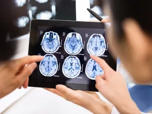
Imagine being able to see inflammation in the brain the way you spot a traffic jam on a live map – instantly, clearly, and without any guesswork. That’s what modern molecular imaging inflammation does with PET scans. In just a few minutes you get a functional snapshot of immune activity, helping doctors diagnose, track, and treat neuro‑inflammatory disorders far earlier than traditional MRI or CT ever could.
So, if you’ve ever wondered whether there’s a way to peek inside the brain’s immune system, the answer is yes – and it’s getting better every day. Below you’ll find a friendly walk‑through of how PET works, why the newest tracer is such a game‑changer, and what it means for patients, clinicians, and anyone curious about the future of brain health.
What Is Molecular Imaging?
Definition and Difference
“Molecular imaging” sounds fancy, but at its core it’s simply imaging that visualises biology at the molecular level instead of just the shape of organs. Think of it as a camera that captures not only the scenery (the brain’s anatomy) but also the actors on stage (immune cells, enzymes, receptors). Traditional scans like CT or structural MRI tell you where something is; molecular imaging tells you what it’s doing.
How PET Works
Positron Emission Tomography (PET) uses a tiny amount of a radioactive molecule called a radiotracer. After you’re injected, the tracer travels through the bloodstream and sticks to a specific target – for inflammation, that’s often a protein on activated microglia (the brain’s resident immune cells). The tracer then emits positrons that collide with electrons, producing gamma rays the scanner picks up. The result is a colorful map where hot spots mean “high inflammation activity.”
Key Biomarkers
Scientists have identified several molecular signposts that PET can chase:
- TSPO – a protein that balloons on activated microglia.
- COX‑2 – an enzyme involved in inflammatory pathways.
- CCR2 and P2X7 – newer targets that may reveal even finer details of the immune response.
Each biomarker needs its own specially‑crafted radiotracer, which is why the market keeps evolving with new “PET tracer brain inflammation” agents.
Brain Inflammation PET
New PET Tracer
In 2024 the Society of Nuclear Medicine and Molecular Imaging (SNMMI) highlighted a breakthrough tracer – let’s call it Fluoro‑DPA‑714. Compared with the classic ^18F‑FDG (glucose) tracer, Fluoro‑DPA‑714 shows more than twice the target‑to‑background ratio in early neuroinflammation, giving doctors a crystal‑clear picture of where microglia are firing up.
According to a 2020 study in the Journal of Nuclear Medicine, TSPO‑targeted tracers like this can detect subtle changes months before symptoms appear, especially in Alzheimer’s and Parkinson’s disease.
Clinical Uses
Here’s where the magic meets the bedside:
- Alzheimer’s disease – early detection of microglial activation helps identify patients who may benefit from anti‑inflammatory therapies.
- Multiple sclerosis – PET can differentiate active lesions from scar tissue, guiding treatment escalation.
- Traumatic brain injury – spotting lingering inflammation after a concussion can explain persistent headaches or mood changes.
- Systemic autoimmune disorders – conditions like lupus or sarcoidosis that affect the brain become easier to monitor.
For a deeper dive on how PET visualises these conditions, check out our guide on PET imaging brain inflammation.
Benefits and Risks
Why It Helps
Here are the headline benefits that make clinicians and patients smile:
- Early diagnosis – Catching inflammation before irreversible damage sets in.
- Therapy guidance – See whether a drug is actually calming the immune fire.
- Quantifiable metrics – Standardised Uptake Values (SUVs) give objective numbers you can compare over time.
- Research power – Uniform imaging endpoints accelerate clinical trials.
Safety Considerations
Of course, no technology is without trade‑offs. PET involves a modest radiation dose – roughly 5 mSv for a brain scan, which is comparable to a few months of natural background radiation. For most adults the risk is very low, especially when the scan is medically justified. The bigger practical concern is access: not every hospital carries the new tracer yet, and production requires a cyclotron or nearby radiopharmacy.
If you’re weighing the pros and cons, ask your doctor about dose‑reduction protocols and whether a combined PET/MRI exam could give you the best of both worlds without extra radiation.
Regulatory Landscape
The new tracer is currently navigating the FDA’s Investigational New Drug (IND) pathway, with Phase II trials showing promising safety and efficacy data. In Europe, the EMA has granted “orphan drug” status for its use in rare neuroinflammatory disorders, which should speed up reimbursement discussions.
Industry giants like Siemens Healthineers and GE Healthcare are already positioning their PET platforms to support the tracer, while biotech startups are developing next‑generation ligands that bind to CCR2 or P2X7. This collaborative ecosystem suggests that broader insurance coverage could arrive within the next few years.
Getting Started
Ready to explore whether a PET scan could help you or a loved one? Here’s a simple roadmap:
- Identify the clinical question. Are you trying to confirm early Alzheimer’s, monitor MS activity, or assess post‑concussion inflammation?
- Select the tracer. For general neuroinflammation, the TSPO‑targeted PET tracer brain inflammation is often the first choice; for more specific pathways, ask about CCR2 or P2X7 agents.
- Schedule the scan. Most centers require a short fasting period (no coffee or sugary snacks) and a brief medication review (some anti‑inflammatories can interfere).
- Interpret the images. Radiologists look at SUV ratios; values above 1.5–2.0 typically indicate active inflammation, but thresholds can vary by tracer and disease.
- Integrate findings. Discuss results with a neurologist or rheumatologist to adjust treatment plans, whether that means starting a new disease‑modifying drug or simply monitoring.
For a practical checklist, see our neuroinflammation PET scan guide.
Future Directions
What’s next on the horizon? A few exciting trends are already reshaping the field:
- Hybrid PET/MRI. Combining PET’s molecular insight with MRI’s exquisite anatomy offers simultaneous functional and structural maps, reducing overall scan time.
- AI‑driven quantification. Machine‑learning algorithms can automatically segment hot spots, calculate SUV‑ratios, and even predict disease progression from raw data.
- Theranostic pairs. The same molecule can be labelled with a diagnostic isotope for imaging or a therapeutic one for targeted radio‑therapy – a true “see‑and‑treat” approach.
- New molecular targets. Research into P2X7, folate receptor β, and other immune markers promises even more precise sub‑typing of inflammation.
If you’re curious about how these innovations translate into everyday care, keep an eye on the upcoming radiotracer brain imaging updates – we’ll be covering the latest trials and what they mean for patients.
Conclusion
Molecular imaging inflammation, especially with PET, is turning what once felt like a mystery into a clear, actionable picture of brain health. The newest PET tracer delivers sharper images, earlier detection, and a richer toolbox for clinicians—all while keeping radiation exposure low and safety high. As regulations catch up and AI refines interpretation, we’re moving toward a future where every patient can get personalized, image‑guided care for neuroinflammatory diseases.
Whether you’re a patient seeking answers, a caregiver navigating treatment options, or just a curious mind, remember that the science is evolving every day. Talk to your neurologist or nuclear medicine specialist about whether a PET scan fits your situation, and stay tuned for the next wave of breakthroughs. After all, seeing is believing – and in the world of brain inflammation, seeing has never been more powerful.


Leave a Reply
You must be logged in to post a comment.